. 2012 Jan;32(1):103-9.
doi: 10.1161/ATVBAHA.111.236349. Epub 2011 Oct 27.

Aging enhances the basal production of IL-6 and CCL2 in vascular smooth muscle cells

Affiliations

Aging enhances the basal production of IL-6 and CCL2 in vascular smooth muscle cells

Yang Song et al. Arterioscler Thromb Vasc Biol. 2012 Jan.

Abstract

Objective: Increased circulating cytokine levels are a prominent feature of aging that may contribute to atherosclerosis. However, the role vascular cells play in chronic inflammation induced by aging is not clear. Here, we examined the role of aging on inflammatory responses of vascular cells.

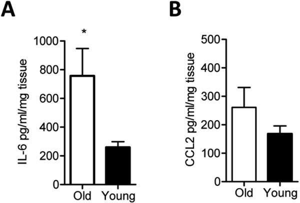
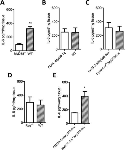
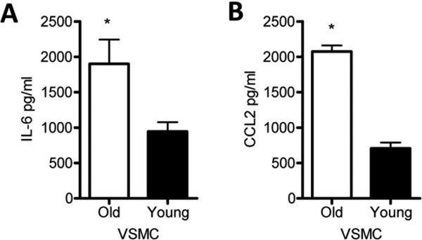
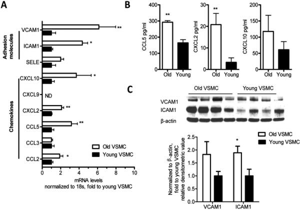
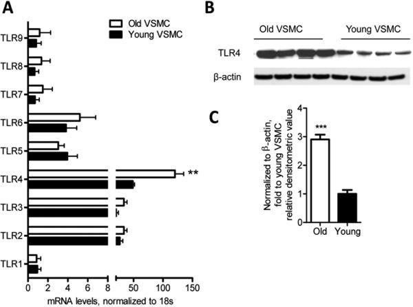
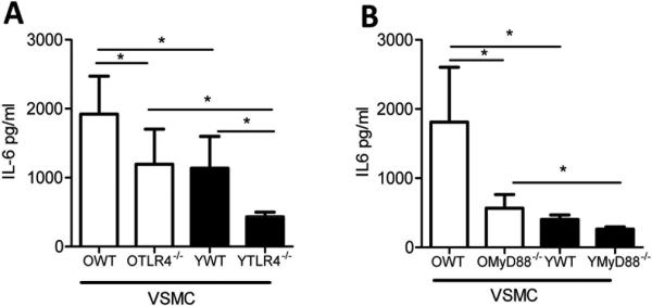
Methods and results: In an ex vivo culture system, we examined the inflammatory response of aortas from young (2-4 months) and aged (16-18 months) mice under nonstimulatory conditions. We found that basal levels of interleukin-6 were increased in aged aortas. Aged aortic vascular smooth muscle cells (VSMC) exhibited a higher basal secretion of interleukin-6 than young VSMC. Gene and protein expression analysis revealed that aged VSMC exhibited upregulation of chemokines (eg, CCL2), adhesion molecules (eg, intracellular adhesion molecule 1), and innate immune receptors (eg, Toll-like receptor [TLR] 4), which all contribute to atherosclerosis. Using VSMC from aged TL4(-/-) and Myd88(-/-) mice, we demonstrate that signaling via TLR4 and its signal adaptor, MyD88, are in part responsible for the age-elevated basal interleukin-6 response.

Conclusions: Aging induces a proinflammatory phenotype in VSMC due in part to increased signaling of TLR4 and MyD88. Our results provide a potential explanation as to why aging leads to chronic inflammation and enhanced atherosclerosis.


Figures

Figure 1
Figure 1. Basal IL-6 cytokine production is increased in aged aortas than young aortas
Thoracic aortas from young or aged mice were harvested and cultured in media devoid of LPS for 16 hours. IL-6 (A) and CCL2 (B) levels were measured by ELISA. The data shown are representative of three independent experiments. Results are presented as means ± SEM, n at least 5 mice / group / experiment. Differences between groups were analyzed using Student's t-test. *, P<0.05. O: old 16-18 month; Y: young 6-8 weeks.
Figure 2
Figure 2. Vascular smooth muscle cells are major contributors to the basal IL-6 response of young murine aortas
Aortas from genetically altered young mice (6-8 weeks of age) were used to identify the MyD88 expressing cells that contribute to the basal IL-6 response. Aortas were harvested from young wild type and Myd88-/- mice and IL-6 measured in the culture supernatants (A). CD11c-Myd88 Tg mice are Myd88-/- except in CD11c+ cells and were compared to wild type controls (B). The aortas of LysMCre/MyD88-flox mice, which are Myd88-/- in Lysm+ cells, were compared to their LysM-Cre-/-/MyD88-flox Cre negative control (C). Rag-/- mice lack mature T cells and B cells and aortas of these mice were compared to wild type controls (D). SM22α-Cre/MyD88-flox are MyD88 deficient in smooth muscle cells and were compared with their Cre negative control (E). IL-6 levels were measured by ELISA. Differences between groups were analyzed using Student's t-test, n = at least 4 mice/group/experiment. *, P<0.05; **, P<0.01. The data shown are representative of three independent experiments.
Figure 3
Figure 3. IL-6 and CCL2 levels were increased in aged VSMC than young VSMC without stimulation
VSMC were isolated from thoracic aortas of young or aged mice as described in Methods, and equal number of cells from young and aged mice were cultured in fresh medium for 16 hours. Basal IL-6 (A) and CCL2 (B) levels measured in culture supernatants were measured by ELISA. The data shown are representative of three independent experiments. Results are presented as means ± SEM, n at least 4 mice/group/experiment as a source of cells. Differences between groups were analyzed using Student's t-test. *, P<0.05. O, old 16-18 month; Y, young 6-8 weeks.
Figure 4
Figure 4. Pro-atherogenic chemokines and adhesion molecules are upregulated in non stimulated VSMC with aging
VSMC were isolated from murine thoracic aortas as described in Methods. Non-stimulated VSMC were collected, and chemokines and adhesion molecules were measured by qRT-PCR (A). Results are presented as means ± SEM. Data shown represent one experiment with n =12 mice as a source of cells / group. CCL5, CXCL2, and CXCL10 were measured in the supernatants of cultured, non-stimulated VSMC by ELISA (B). Western blot analysis measuring ICAM1 and VCAM1 protein levels (C). Differences between young and aged VSMC were analyzed using Student's t-test. *, P<0.05; **P<0.01. In (C) the values are normalized to β-actin expression and represent fold change to young VSMC value. Data shown in (B) and (C) are representative of two independent experiments, with n = at least 4 mice / group / experiment as a source of cells. All comparisons between young and aged groups were paired and performed contemporaneously.
Figure 5
Figure 5. VSMC express increased TLR4 gene expression and protein levels with aging
TLR gene expression profiles were evaluated in aged and young non-stimulated VSMC by qRT-PCR. TLR4 expression was significantly higher in aged VSMC than young VSMC (A). Western blot analysis showing that aged VSMC exhibit higher TLR4 protein levels than young VSMC (B-C). Results are presented as means ± SEM and in (A) data shown represent one experiment with n = 15 mice as a source of cells / group. In (C) the values are normalized to β-actin expression and represent fold change relative to young VSMC value. In (B) and (C), data shown are representative of two independent experiments, with n = 4 mice / group / experiment as a source of cells. Differences between young and aged VSMC were analyzed using Student's t-test. **, P<0.01, ***, P<0.0001. All comparisons between young and aged groups were paired and performed contemporaneously.
Figure 6
Figure 6. Elevated IL-6 secretion by aged VSMC and aortas is in part TLR4 and MyD88 dependent
Aortas were harvested from aged (10-11 month of age) or young (6-8 weeks of age) WT, TLR4-/- or Myd88-/- mice, and VSMC were isolated from these aortas. VSMC were cultured in fresh medium for 16 hours without stimulation, and IL-6 was measured in the supernatants by ELISA. Aged TLR4-/- (denoted as OTLR4-/-) (A) Myd88-/- (denoted as OMyD88-/-) (B) VSMC exhibited a significant reduction in IL-6 production in comparison with the aged WT counterparts. This was also evident in the young VSMC. Results are presented as means ± SD, n = 3 mice / group for both panels A and B. Differences between groups were analyzed using one-way ANOVA with Bonferroni's post-hoc test. *, P<0.05. O:old 10-11 month; Y: young 6-8 weeks.

References

    1. Collaboration Asia Pacific Cohort Studies Collaboration The impact of cardiovascular risk factors on the age-related excess risk of coronary heart disease. Int J Epidemiol. 2006;35:1025–1033. - PubMed
    1. Robak T, Gladalska A, Stepien H, Robak E. Serum levels of interleukin-6 type cytokines and soluble interleukin-6 receptor in patients with rheumatoid arthritis. Mediators Inflamm. 1998;7:347–353. - PMC - PubMed
    1. Robak E, Sysa-Jedrzejowska A, Stepien H, Robak T. Circulating interleukin-6 type cytokines in patients with systemic lupus erythematosus. Eur Cytokine Netw. 1997;8:281–286. - PubMed
    1. Deeva I, Mariani S, De Luca C, Pacifico V, Leoni L, Raskovic D, Kharaeva Z, Korkina L, Pastore S. Wide-spectrum profile of inflammatory mediators in the plasma and scales of patients with psoriatic disease. Cytokine. 2010;49:163–170. - PubMed
    1. McMahon M, Hahn BH, Skaggs BJ. Systemic lupus erythematosus and cardiovascular disease: Prediction and potential for therapeutic intervention. Expert Rev Clin Immunol. 2011;7:227–241. - PMC - PubMed

Publication types

MeSH terms