. 2011 Dec;59(12):1087-100.
doi: 10.1369/0022155411423680. Epub 2011 Oct 27.

Human breast cancer histoid: an in vitro 3-dimensional co-culture model that mimics breast cancer tissue

Affiliations

Human breast cancer histoid: an in vitro 3-dimensional co-culture model that mimics breast cancer tissue

Pavinder Kaur et al. J Histochem Cytochem. 2011 Dec.

Abstract

Progress in our understanding of heterotypic cellular interaction in the tumor microenvironment, which is recognized to play major roles in cancer progression, has been hampered due to unavailability of an appropriate in vitro co-culture model. The aim of this study was to generate an in vitro 3-dimensional human breast cancer model, which consists of cancer cells and fibroblasts. Breast cancer cells (UACC-893) and fibroblasts at various densities were co-cultured in a rotating suspension culture system to establish co-culture parameters. Subsequently, UACC-893, BT.20, or MDA.MB.453 were co-cultured with fibroblasts for 9 days. Co-cultures resulted in the generation of breast cancer histoid (BCH) with cancer cells showing the invasion of fibroblast spheroids, which were visualized by immunohistochemical (IHC) staining of sections (4 µm thick) of BCH. A reproducible quantitative expression of C-erbB.2 was detected in UACC-893 cancer cells in BCH sections by IHC staining and the Automated Cellular Imaging System. BCH sections also consistently exhibited qualitative expression of pancytokeratins, p53, Ki-67, or E-cadherin in cancer cells and that of vimentin or GSTPi in fibroblasts, fibronectin in the basement membrane and collagen IV in the extracellular matrix. The expression of the protein analytes and cellular architecture of BCH were markedly similar to those of breast cancer tissue.


Conflict of interest statement

The authors declared no potential conflicts of interest with respect to the authorship and publication of this article.

Figures

Figure 1.
Figure 1.
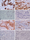
Evaluation of UACC-893 breast cancer histoid (BCH). The sections (4-µm thick) of formalin-fixed and paraffin-embedded BCH, resulting from a co-culture of the human breast cancer cell line UACC-893 (1 × 10⁶) and foreskin fibroblasts (FSF) (2 × 10⁷) in the bioreactor for 1, 2, or 9 days, were immunohistochemically stained with mouse monoclonal antibodies to pancytokeratins or vimentin. A representative example of pancytokeratin-positive cancer cells (reddish brown staining, indicated by closed arrows) shows coating of the external layer of an individual pancytokeratin-negative FSF spheroid (indicated by open arrows) at the first 24 hours of co-culture (A). In a consecutive section, an individual FSF spheroid shows reactivity with anti-vimentin antibody (reddish brown staining, indicated by open arrows), whereas the surrounding cancer cells were nonreactive (absence of reddish brown staining, indicated by closed arrows) (B). The pancytokeratin-positive cancer cells (indicated by closed arrows) showed invasion of the core of an individual FSF spheroid (indicated by open arrows) at 48 hours in co-culture (C). Process of invasion of the FSF spheroid by the cancer cells was complete in 9 days in co-culture, as most of the pancytokeratin-positive cancer cells (closed arrows) were detected within the core of an individual FSF spheroid (open arrows) (D). The sections were counterstained with hematoxylin (blue nuclear staining). Original magnification: A–C (×200) and D (×120). Bar = 100 µm.
Figure 2.
Figure 2.
Immunohistochemical localization of C-erbB.2 protein expression in UACC-893 breast cancer histoid (BCH) and breast cancer tissue. The sections (4-µm thick) of formalin-fixed and paraffin-embedded BCH preparation (A, C), resulting from co-culture of breast cancer cell line UACC-893 (1 × 10⁶) and fibroblast (FSF) (2 × 10⁷) in the bioreactor for 9 days, or human breast cancer tissue (B, D) were immunohistochemically stained with rabbit polyclonal antibody to the C-erbB.2 protein. The antibody exhibited membranous staining of the cancer cells, as indicated by reddish brown staining (closed arrows) in BCH (A) or breast cancer tissue (B), whereas the fibroblasts, as indicated by the absence of reddish brown staining (open arrows), showed no reactivity with the antibody in BCH (A) or breast cancer tissue (B). The application of preabsorbed anti-C-erbB.2 antibody with specific blocking peptide led to abolition of immunostaining of the target cells, as indicated by the absence of reddish brown staining (closed arrow) in BCH (C) or breast cancer tissue (D). The sections were counterstained with hematoxylin (blue nuclear staining). Original magnification: ×310. Bar = 100 µm.
Figure 3.
Figure 3.
Quantitative intensity of image analysis of immunochemical expression of the C-erbB.2 protein in the UACC-893 human breast cancer cell line in breast cancer histoid (BCH) by the Automated Cellular Imaging System III (ACIS III). Each block of BCH was obtained from a co-culture of breast cancer cell line (1 × 10⁶) and FSF (2 × 10⁷) for 9 days. Quantitative intensity of image of C-erbB.2 protein expression was analyzed by ACIS III on C-erbB.2–immunostained sections, representing every fifth section from a total of 75 sections from two of three formalin-fixed and paraffin-embedded BCH blocks from each of the three separate batches of BCH preparations (16 sections per block × 6 = 96 sections). The ACIS III quantitates images on a scale of 0 to 5. The bars represent standard error of immunohistochemical expression of C-erbB.2 within the six randomly selected BCH constructs per section. The quantitative expression of C-erbB.2 in the cancer cells in 16 sections from each of the six BCH preparations (blocks) showed scores that ranged from 3.1 to 5.0 (standard error = 0.03). The coefficient of variation (CV) was 4.21 for the average score. No statistically significant difference in the score was observed between the sections from intrabatches or interbatches of BCH preparations (p=0.61).
Figure 4.
Figure 4.
Immunohistochemical comparison of the expression of breast tissue–associated protein analytes between UACC-893 breast cancer histoid (BCH) and breast cancer tissue. The sections (4-µm thick) of formalin-fixed and paraffin-embedded BCH (A, C, E, G), resulting from a co-culture of a human breast cancer cell line (UACC-893) and fibroblasts (FSF) in the bioreactor for 9 days, or human breast cancer tissue (B, D, F, H) were immunohistochemically stained. The anti-pancytokeratin antibodies showed cytoplasmic reactivity with the cancer cells, as indicated by the reddish brown staining (closed arrow), in BCH (A) or breast cancer tissue (B), whereas the fibroblasts were nonreactive, as indicated by the absence of reddish brown staining (open arrow) (A, B). The application of preabsorbed anti-pancytokeratin antibodies led to the abolition of immunostaining of the target cells, as indicated by the absence of reddish brown staining (closed arrows), in BCH (C) or breast cancer tissue (D). The anti-vimentin antibody showed cytoplasmic reactivity with the fibroblasts, as indicated by reddish brown staining (closed arrows), in BCH (E) and breast cancer tissue (F), whereas the cancer cells were negative, indicated by the absence of reddish brown staining (open arrows) (E, F). The application of preabsorbed anti-vimentin antibody with specific blocking peptide led to the abolition of immunostaining of the target cells, as indicated by the absence of reddish brown staining (closed arrows), in BCH (G) or breast cancer tissue (H). The sections were counterstained with hematoxylin (blue nuclear staining). Original magnification: ×310. Bar = 100 µm.
Figure 5.
Figure 5.
Immunohistochemical comparison of the expression of breast cancer cells, fibroblast, basement membrane, or extracellular matrix–associated protein analytes between UACC-893 breast cancer histoid (BCH) and breast cancer tissue. The sections (4-µm thick) of formalin-fixed and paraffin-embedded BCH (A, C, E, G, I, K), resulting from a co-culture of a human breast cancer cell line (UACC-893) and fibroblasts (FSF) in the bioreactor for 9 days, or human breast cancer tissue (B, D, F, H, J, L) were immunohistochemically stained. The antibodies to p53 or Ki-67 exhibited nuclear reactivity with the cancer cells, as indicated by reddish brown staining (open arrows), in BCH (A, C) or breast cancer tissue (B, D), whereas the fibroblasts were nonreactive with the antibody, as indicated by the absence of reddish brown staining (open arrows), in BCH (A, C) or breast cancer tissue (B, D). Bar = 100 µm. Antibody to E-cadherin showed membranous reactivity with the cancer cells in BCH (E) and breast cancer tissue (F). Bar = 100 µm. Anti-GSTPi antibody showed cytoplasmic/nuclear reactivity with the fibroblasts, as indicated by brown staining (open arrows), in BCH (G) or breast cancer tissue (H), whereas the cancer cells were nonreactive, as indicated by the absence of brown staining (closed arrows), in BCH (G) or breast cancer tissue (H). The anti-fibronectin antibody exhibited reactivity with the basement membrane component, as indicated by reddish brown staining (closed arrows), in BCH (I) or breast cancer tissue (J). The anti–collagen IV antibody showed reactivity with the extracellular matrix, as indicated by reddish brown staining (closed arrows), in BCH (K) or breast cancer tissue (L). The sections were counterstained with hematoxylin (blue nuclear staining). Original magnification: A–D and G–L (×310), E (×150), and F (×210). Bar = 100 µm.
Figure 6.
Figure 6.
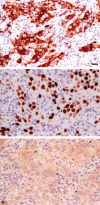
Immunohistochemical expression of breast tissue–associated protein analytes in BT.20 breast cancer histoid (BCH). The sections (4-µm thick) of formalin-fixed and paraffin-embedded BCH, resulting from a co-culture of the BT.20 breast cancer cell line and fibroblasts (FSF) in the bioreactor for 9 days, were immunohistochemically stained. Antibodies to pancytokeratins or Ki-67 exhibited cytoplasmic or nuclear reactivity, respectively, as indicated by the reddish brown staining with BT.20 breast cancer cells in BCH (A, B, respectively) (closed arrow), whereas fibroblasts were nonreactive, as indicated by the absence of reddish brown staining with both antibodies (A, B) (open arrows). Antibody to C-erbB.2 exhibited a weak cytoplasmic reactivity with BT.20 breast cancer cells in BCH (C) (closed arrow), whereas the fibroblasts were nonreactive, as indicated by open arrows. The sections were counterstained with hematoxylin (blue nuclear staining). Original magnification: ×310. Bar = 100 µm.

References

    1. Bauer M, Su G, Beebe D, Friedl A. 2010. 3D microchannel co-culture: method and biological validation. Integr Biol. 2:371–378 - PMC - PubMed
    1. Beliveau A, Mott JD, Lo A, et al. 2011. Raf-induced MMP9 disrupts tissue architecture of human breast cells in three-dimensional culture and is necessary for tumor growth in vivo. Genes Dev. 24:2800–2811 - PMC - PubMed
    1. Burstein HJ, Chen Y-H, Parker LM, et al. 2008. VEGF as a marker for outcome among advanced breast cancer patients receiving anti-VEGF therapy with bevacizumab and vinorelbine chemotherapy. Clin Cancer Res. 14:7871–7877 - PubMed
    1. Che ZM, Jung TH, Choi JH, et al. 2006. Collagen-based co-culture for invasive study on cancer cells-fibroblasts interaction. Biochem Biophys Res Commun. 346:268–275 - PubMed
    1. Chung LW, Baseman A, Assikis V, et al. 2005. Molecular insights into prostate cancer progression: the missing link of tumor microenvironment. J Urol. 173:10–20 - PubMed

Publication types

Substances