. 2011 Jan-Mar;1(1):17-32.
doi: 10.4103/2045-8932.78097.

Protein trafficking dysfunctions: Role in the pathogenesis of pulmonary arterial hypertension

Affiliations

Protein trafficking dysfunctions: Role in the pathogenesis of pulmonary arterial hypertension

Pravin B Sehgal et al. Pulm Circ. 2011 Jan-Mar.

Abstract

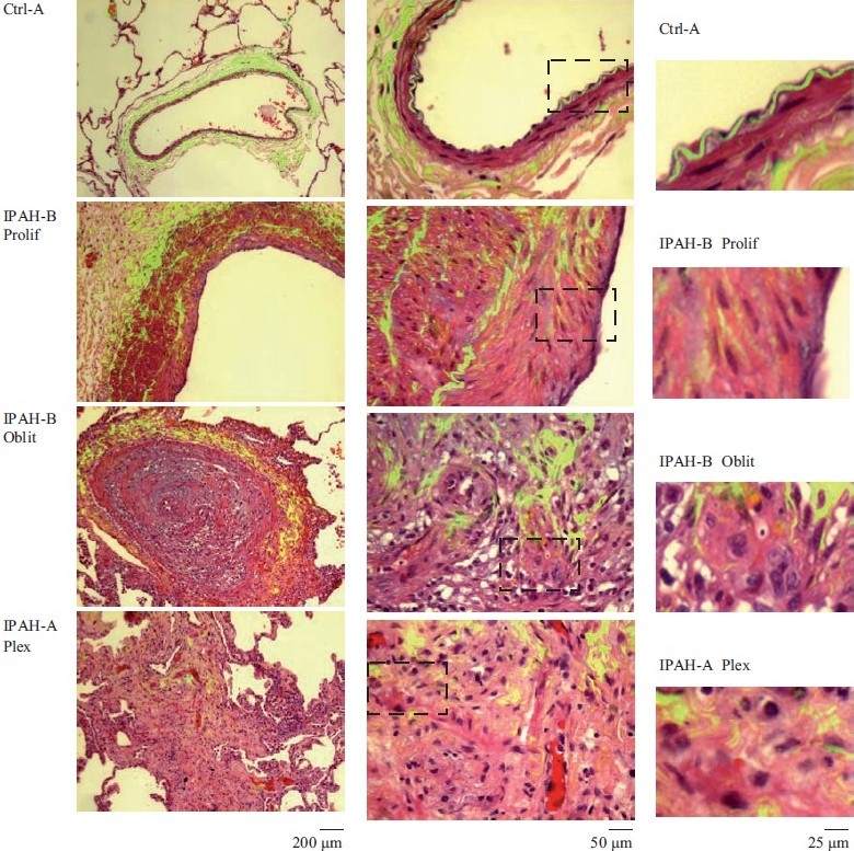
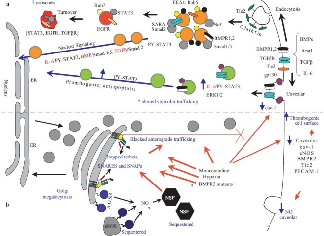
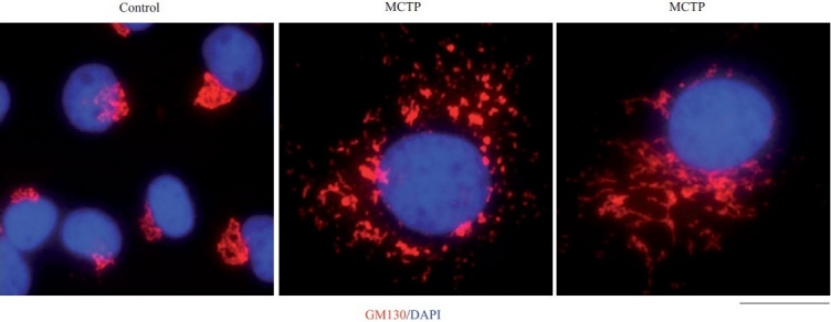
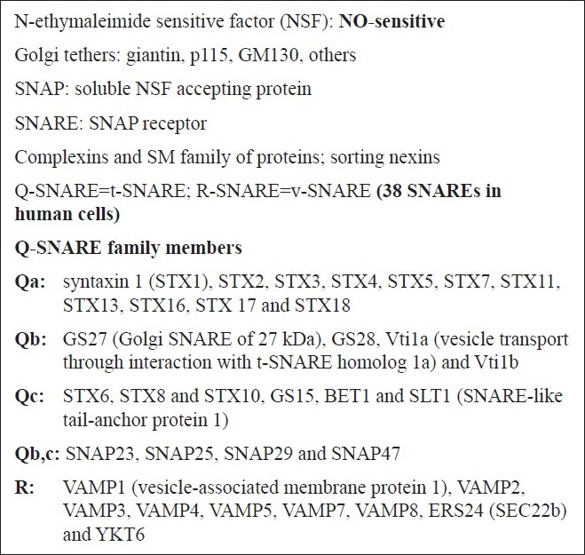
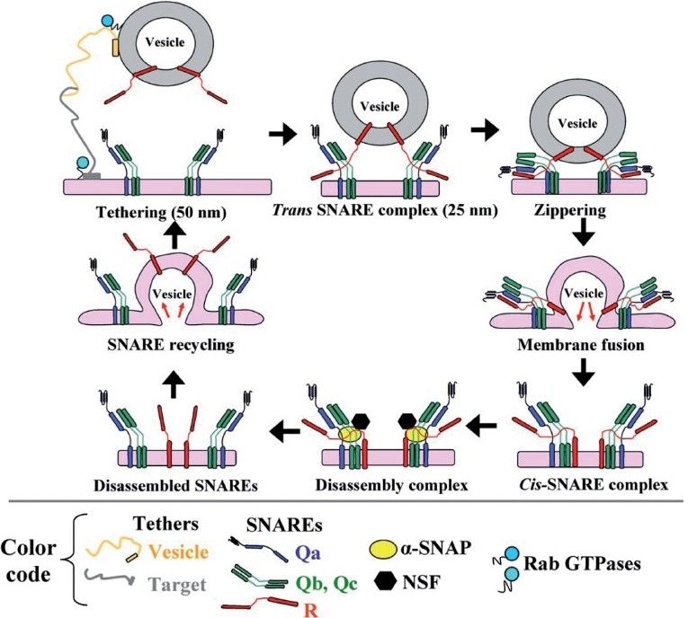
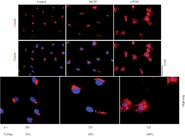
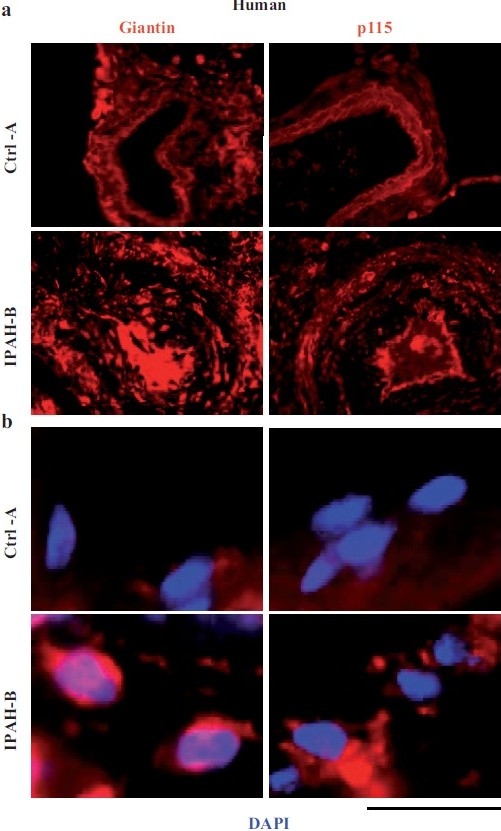
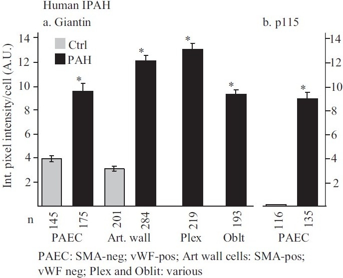
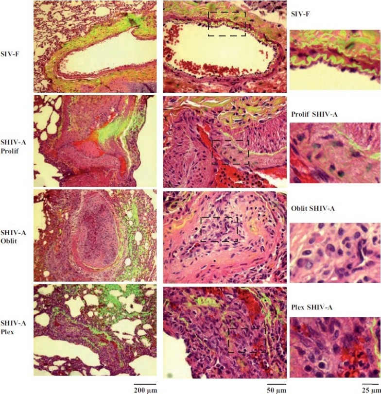
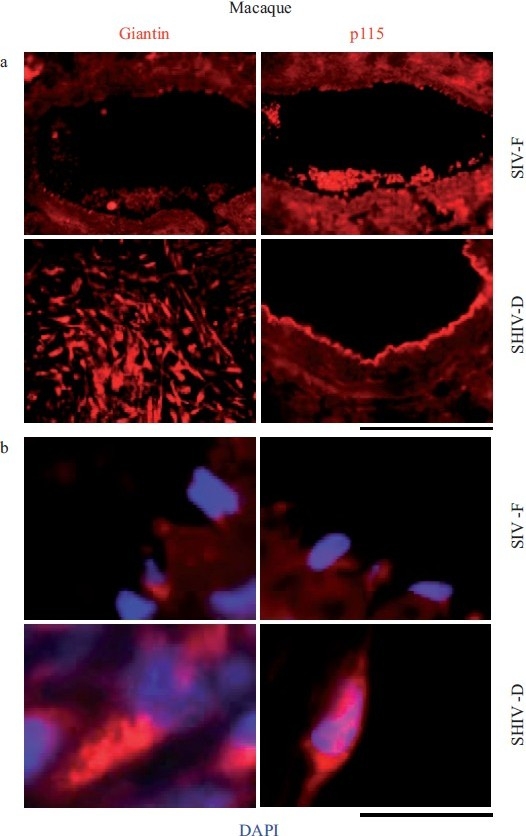
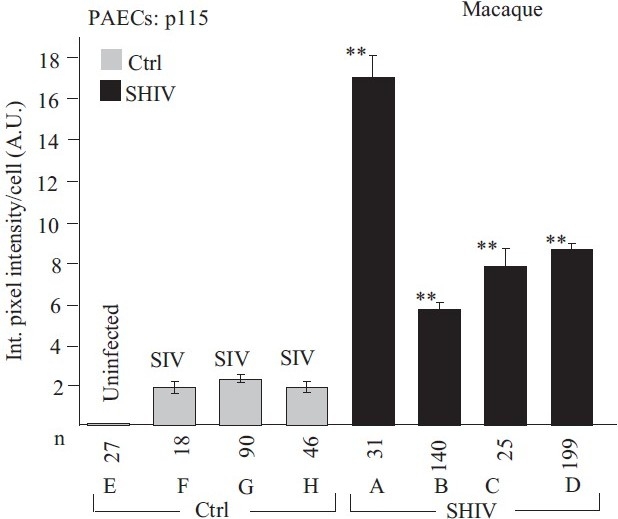
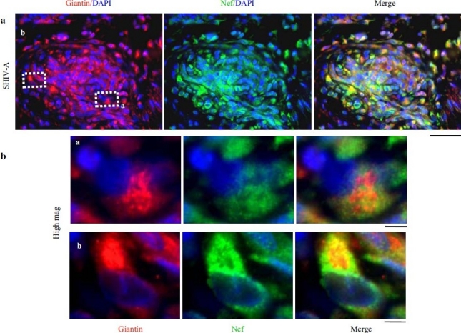
Earlier electron microscopic data had shown that a hallmark of the vascular remodeling in pulmonary arterial hypertension (PAH) in man and experimental models includes enlarged vacuolated endothelial and smooth muscle cells with increased endoplasmic reticulum and Golgi stacks in pulmonary arterial lesions. In cell culture and in vivo experiments in the monocrotaline model, we observed disruption of Golgi function and intracellular trafficking with trapping of diverse vesicle tethers, SNAREs and SNAPs in the Golgi membranes of enlarged pulmonary arterial endothelial cells (PAECs) and pulmonary arterial smooth muscle cells (PASMCs). Consequences included the loss of cell surface caveolin-1, hyperactivation of STAT3, mislocalization of eNOS with reduced cell surface/caveolar NO and hypo-S-nitrosylation of trafficking-relevant proteins. Similar Golgi tether, SNARE and SNAP dysfunctions were also observed in hypoxic PAECs in culture and in PAECs subjected to NO scavenging. Strikingly, a hypo-NO state promoted PAEC mitosis and cell proliferation. Golgi dysfunction was also observed in pulmonary vascular cells in idiopathic PAH (IPAH) in terms of a marked cytoplasmic dispersal and increased cellular content of the Golgi tethers, giantin and p115, in cells in the proliferative, obliterative and plexiform lesions in IPAH. The question of whether there might be a causal relationship between trafficking dysfunction and vasculopathies of PAH was approached by genetic means using HIV-nef, a protein that disrupts endocytic and trans-Golgi trafficking. Macaques infected with a chimeric simian immunodeficiency virus (SIV) containing the HIV-nef gene (SHIV-nef), but not the non-chimeric SIV virus containing the endogenous SIV-nef gene, displayed pulmonary arterial vasculopathies similar to those in human IPAH. Only macaques infected with chimeric SHIV-nef showed pulmonary vascular lesions containing cells with dramatic cytoplasmic dispersal and increase in giantin and p115. Specifically, it was the HIV-nef-positive cells that showed increased giantin. Elucidating how each of these changes fits into the multifactorial context of hypoxia, reduced NO bioavailability, mutations in BMPR II, modulation of disease penetrance and gender effects in disease occurrence in the pathogenesis of PAH is part of the road ahead.

Keywords: Golgi apparatus; Pulmonary vascular remodeling; intracellular organelles.


Conflict of interest statement

Conflict of Interest: None

Figures

Figure 1
Figure 1
Representative histopathologic changes observed in idiopathic pulmonary hypertension. Sections of human lungs (Ctrl-A, IPAH-A and IPAH-B) were stained using H&E and imaged using a ×40 objective in visible light. Elastin autofl uoresence was simultaneously imaged in green and the visible light and autofluoresence images merged. Representative images showing neointimal proliferation (Prolif), obliterative (Oblit) and plexiform (Plex) lesions are illustrated. Side sets on the right show higher magnifi cation views of the boxed areas within panels in the middle column. (Adapted from ref. 2.)
Figure 2
Figure 2
Dysfunctional intracellular trafficking in the pathobiology of pulmonary arterial hypertension. (a) Productive transcriptional signaling from the plasma membrane to the nucleus along the BMP/Smad1/5, TGFβ/Smad2 and IL-6/PY-STAT-3 signaling pathways is membrane associated. IL-6/STAT3 and ERK1/2 signaling is inversely related to loss of caveolar/raft cav-1. (b) Golgi blockade mechanisms in PAH. MCTP and hypoxia lead to a trapping of vesicle tethers, SNAREs and SNAPs in the Golgi of affected pulmonary arterial endothelial cells. This leads to a block in anterograde trafficking of vasorelevant cargo proteins such as cav-1 and eNOS and reduced caveolar NO production. The intracellularly sequestered eNOS produces NO which may potentially S-nitrosylate cysteine-rich proteins like NSF, further inhibiting traffi cking. Golgi-trapped dominant negative BMPR 2 mutants may also potentially block trafficking of cargo proteins to the plasma membrane. (Adapted from ref. 8.)
Figure 3
Figure 3
Golgi enlargement and fragmentation in primary bovine PAECs in culture exposed to MCTP for 4 days. BPAEC cultures in a 6-well plate were exposed to MCTP and megalocytosis was allowed to develop for 4 days.[378] The cultures were then fixed and immunostained for the Golgi tether, GM130, and for nuclei using 4’,6-diamidino-2-phenylindole (DAPI). Scale bar=4 μm.
Figure 4
Figure 4
Tethers, SNAREs, SNAPs, NSF and additional molecules involved in vesicular trafficking
Figure 5
Figure 5
The SNARE cycle in membrane fusion. Initial interaction between a vesicle and its target membrane is mediated by cognate tethers. Membrane fusion is then mediated by the formation of a quaternary-α-helical trans-SNARE complex consisting of one v- (or R -) SNARE on the vesicle and two or three t- (or Q-) SNAREs on the target membrane. After membrane fusion, the cis-SNARE complex is disassembled by the ATPase NSF which is recruited to the cis-SNARE complex from the cytosol by α-SNAP. (Adapted from ref. 8.)
Figure 6
Figure 6
Golgi fragmentation and dispersal in human PAECs after exposure to MCTP or the NO scavenger, c-PTIO. Primary HPAEC cultures in 6-well plates were exposed to MCTP once or to c-PTIO (100 μM) continuously for 48 hrs. The integrity of the Golgi was assayed by immunotagging for the Golgi tether, giantin, together with DAPI staining for demarcating nuclei. NIH Image J software using Otsu segmentation analyses were used to determine Golgi structures with fragment number greater than 1[75] and are enumerated as % cells with dispersed Golgi. Scale bar=20 μm
Figure 7
Figure 7
Increased accumulation of the Golgi matrix proteins/tethers, giantin and p115, in cellular elements in pulmonary arterial vasculopathies in human IPAH. (a) Representative images of respective vasculopathies probed for giantin or p115 compared to representative controls. Scale bar=85 μm. (b) Representative higher magnifi cation images of giantin and p115 immunostaining from analyses as in Figure 7a. Scale bar=10 μm. (Adapted from ref. 2.)
Figure 8
Figure 8
Summary of the quantitative immunomorphometry data for the Golgi tethers, giantin and p115, in cellular elements in pulmonary arterial vasculopathies in IPAH. Integrated pixel intensity/cell was computed using Image J software by outlining individual cells within immunofluorescence images corresponding to luminal endothelium (PAEC), cells in arterial walls, in plexiform lesions and in obliterative lesions and was expressed in arbitrary units (A.U.). Images for quantitation were derived from sections in the set of the human control and IPAH patients as in ref. 2. All the per cell data (n as indicated in the figure) were pooled into either control or IPAH and evaluated using Student's two-tailed t-test. Asterisks indicate P<0.0001 for groups compared with the corresponding control groups; cells in plexiform lesions and in the obliterative lesions were compared with the control PAEC group. In Panel b, there was little detectable p115 signal in the control PAECs evaluated. (Adapted from ref. 2.)
Figure 9
Figure 9
Representative histopathologic changes observed in pulmonary vascular lesions in SHIV-nef–infected macaques. Sections of lungs from macaquesinfected with the chimeric SHIV-nef virus (SHIV-A) or a non-chimeric SIV virus (SIV-F) were stained using H&E and imaged using a ×40 objective in visiblelight. Elastin autofl uoresence was simultaneously imaged in green and the visible light and autofl uoresence images merged. Representative images showing neointimal proliferation (Prolif), obliterative (Oblit) and plexiform (Plex) lesions are illustrated. Side sets on the right show higher magnifi cation views of the boxed areas within each panel in the middle column. (Adapted from ref. 2.)
Figure 10
Figure 10
Increased accumulation of the Golgi matrix proteins/tethers, giantin and p115, in cellular elements in pulmonary arterial vasculopathies in the chimeric SHIV-infected macaque model. (a) Representative images of respective vasculopathies probed for giantin or p115 compared to representative controls. Scale bar=85 μm. (b) Representative higher magnification images of giantin and p115 immunostaining from analyses as in Figure 10a. Scale bar=10 μm. The SIV-F, p115 segment in the upper right is the same as in Figure 9, SIV-F at the top of the middle column. (Adapted from ref. 2.)
Figure 11
Figure 11
Quantitative immunomorphometry data for the Golgi tether, p115, in PAECs in SHIV-nef–infected macaques. Integrated pixel intensity/cell was computed using Image J software by outlining individual cells within immunofl uorescence images corresponding to luminal endothelium (PAEC) in sections from each of the control/SIV- and SHIV-infected macaques (n=4 in each group) and was expressed in arbitrary units (A.U.) (mean±SE; n is number of cells quantitated). PAECs in group Ctrl-E had little or no detectable p115. Post-hoc between-group comparisons were carried out using the Tukey–Kramer Multiple Comparison test with an alpha setting of 0.05 (NCSS 2007). Double asterisks indicate that the particular group was different from all control/SIV groups. (Adapted from ref. 2.)
Figure 12
Figure 12
HIV-nef–positive vascular cell elements have increased giantin. (a) Lung sections from a SHIV-nef–infected macaque with increased giantin in the obliterative vascular lesion probed for HIV-nef and giantin. Scale bar=25 μm. (b) Higher magnifi cation images of the insets depicted in Figure 12a. Scale bar=4μm. Respective 3D intracellular immunoimaging is in ref. 75. (Adapted from ref. 2.)

References

    1. Stenmark KR, Meyrick B, Galie N, Mooi WJ, McMurtry IF. Animal models of pulmonary arterial hypertension: The hope for etiological discovery and pharmacological cure. Am J Physiol Lung Cell Mol Physiol. 2009;297:L1013–32. - PubMed
    1. Sehgal PB, Mukhopadhyay S, Patel K, Xu F, Almodóvar S, Tuder RM, et al. Golgi dysfunction is a common feature in idiopathic human pulmonary hypertension and vascular lesions in SHIV-nef-infected macaques. Am J Physiol Lung Cell Mol Physiol. 2009;297:L729–37. - PMC - PubMed
    1. Mathew R, Huang J, Shah M, Patel K, Gewitz M, Sehgal PB. Disruption of endothelial-cell caveolin-1alpha/raft scaffolding during development of monocrotaline-induced pulmonary hypertension. Circulation. 2004;110:1499–506. - PubMed
    1. Zhao YY, Lui Y, Stan RV, Fan L, Gu Y, Dalton N, et al. Defects in caveolin-1 cause dilated cardiomyopathy and pulmonary hypertension in knockout mice. Proc Natl Acad Sci U S A. 2002;99:11375–80. - PMC - PubMed
    1. Zhao YY, Zhao YD, Mirza MK, Huang JH, Potula HH, Vogel SM, et al. Persistent eNOS activation secondary to caveolin-1 deficiency induces pulmonary hypertension in mice and humans through PKG nitration. J Clin Invest. 2009;119:2009–18. - PMC - PubMed

LinkOut - more resources