. 2012 Jan;23(1):155-64.
doi: 10.1681/ASN.2010080858. Epub 2011 Oct 27.

Elevated soluble Flt1 inhibits endothelial repair in PR3-ANCA-associated vasculitis

Affiliations

Elevated soluble Flt1 inhibits endothelial repair in PR3-ANCA-associated vasculitis

Sandrine Le Roux et al. J Am Soc Nephrol. 2012 Jan.

Abstract

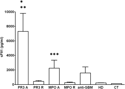
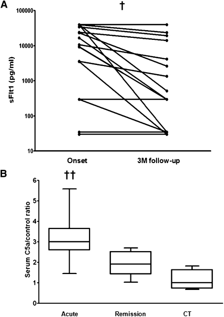
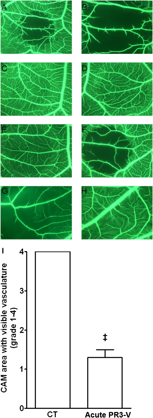
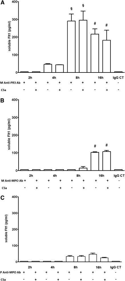
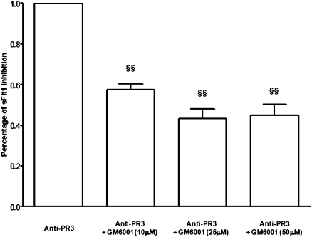
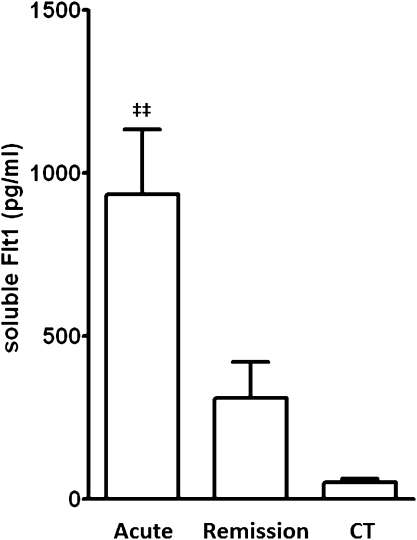
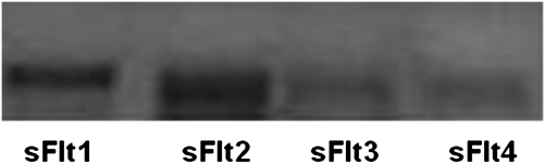
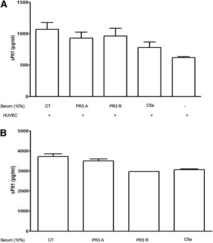
Antineutrophil cytoplasmic antibody (ANCA)-associated vasculitis exhibits endothelial damage, but the capacity for vessel repair in this disorder is not well understood. Here, we observed a marked increase in serum levels of soluble Flt1 (sFlt1), a potent inhibitor of vascular endothelial growth factor, in patients with active ANCA-associated vasculitis compared with patients during remission and other controls. Serum levels of sFlt1 correlated with C5a, an anaphylatoxin released after complement activation. Serum from patients with acute ANCA-associated vasculitis disrupted blood flow in the chicken chorioallantoic membrane assay, suggesting an antiangiogenic effect. Preincubation with excess human vascular endothelial growth factor prevented this effect. Anti-proteinase-3 (PR3) mAb and serum containing PR3-ANCA from patients with active vasculitis both induced a significant and sustained release of sFlt1 from monocytes, whereas anti-myeloperoxidase (MPO) mAb or polyclonal antibodies did not. However, the serum containing polyclonal PR3-ANCA did not induce release of sFlt1 from cultured human umbilical vein endothelial cells. In summary, these data suggest that anti-PR3 antibodies, and to a much lesser extent anti-MPO antibodies, increase sFlt1 during acute ANCA-associated vasculitis, leading to an antiangiogenic state that hinders endothelial repair.


Figures

Figure 1.
Figure 1.
Serum sFlt1 levels in patients with PR3- and MPO-associated vasculitis during acute disease or during disease remission. A, acute disease; CT, healthy controls; GBM, patients with anti-GBM disease; HD, hemodialysis patients; R, remission. *P<0.0001 for PR3 A versus CT, and **P<0.001 for PR3 A versus PR3 R, MPO R, and HD. ***P<0.001 for MPO A versus CT.
Figure 2.
Figure 2.
Serum sFlt1 levels are increased during acute PR3-associated vasculitis. (A) Serum sFlt1 levels (log scale) in 18 patients with PR3-associated vasculitis at the disease onset and at 3-month follow-up. P=0.0026, paired t test. (B) Serum C5a levels in acute and remission ANCA-associated vasculitis samples and from healthy controls demonstrating significantly elevated levels in acute disease compared with disease remission and those found in healthy controls. CT, healthy controls. ††P=0.001 for ANCA-associated vasculitis acute versus remission and controls.
Figure 3.
Figure 3.
Serum from acute PR3-associated vasculitis disrupts blood flow in the CAM microvasculature. Representative pictures of distinct CAM areas treated with the following: (1) serum from a patient with acute PR3-associated vasculitis (A, original magnification, ×31.25 and B, original magnification, ×34); (2) PBS (C, original magnification, ×31.25), (3) healthy control serum (D, original magnification, ×31.25); (4) low molecular (10 kD) (E, original magnification, ×31.25) or high molecular (0.010 kD) (F, original magnification, ×31.25) mass fraction of the serum from a patient with acute PR3-associated vasculitis; and (5) serum from a patient with acute PR3-ANCA–associated vasculitis without (G, original magnification, ×34) or with preincubation (H, original magnification, ×34) with human recombinant VEGF. (I) Semiquantification of the effect of acute anti-PR3–associated vasculitis or control (CT) sera on the microvasculature of the treated CAM area. The effect was graded from 1 to 4 depending on the area of the treated CAM in which microvasculature was still visible 3 hours after the deposition of serum: grade 1 (0%–25% of total treated area), grade 2 (26%–50%), grade 3 (51%–75%), and grade 4 (76%–100%). P<0.0002, paired t test.
Figure 4.
Figure 4.
Monoclonal anti-PR3 antibody induces sFlt1 release from monocytes. (A) An anti-PR3 mAb (10 μg/ml) induced sFlt1 release by human monocytes as early as 4 hours of incubation. sFlt1 release peaked at 8 hours and remained significantly increased at 16 hours. Further addition of C5a (10 nM) for 2 hours did not alter sFlt1 release. Data are from five independent experiments. CT, isotype-matched control. §P<0.001 versus 2-hour, 4-hour, and IgG isotype-matched control (CT) groups. #P<0.01 versus 2-hour, 4-hour, and IgG CT groups. (B) An anti-MPO mAb (10 μg/ml) induced a mild sFlt1 release by human monocytes only after 16 hours of incubation. Further addition of C5a (10 nM) for 2 hours did not alter sFlt1 release. Data are from three to four independent experiments. #P<0.01 versus 2-hour, 4-hour, and IgG CT groups. (C) Polyclonal anti-MPO antibody (10 μg/L) purified from 3 patients with anti-MPO vasculitis induced a very mild increase in sFlt1 release by human monocytes after 8 and 16 hours of incubation. Further addition of C5a (10 nM) for 2 hours did not alter sFlt1 release. Polyclonal anti-MPO IgG from each patient were tested in three independent experiments.
Figure 5.
Figure 5.
An antimetalloprotease inhibitor partially blunted sFlt1 release by monocytes stimulated with anti-PR3 mAb (10 μg/L). Monocytes were pretreated with a metalloprotease inhibitor GM6001 (10, 25, or 50 μM for 1 hour at 37°C) before incubation with anti-PR3 mAb for 16 hours. §§P<0.001 versus anti-PR3.
Figure 6.
Figure 6.
Serum (10%) from 12 patients with PR3-ANCA during acute ANCA-associated vasculitis induced significantly more sFlt1 release by monocytes after 16 hours of incubation, compared with serum from the same patients during disease remission or control serum. CT, control. ‡‡P<0.001 versus all other groups.
Figure 7.
Figure 7.
sFlt1 isoforms 1 and 2 were predominant in monocytes at baseline and after stimulation (1 hour) with anti-PR3 antibodies, whereas isoform 3 and 4 transcripts were barely detectable. A representative result of sFlt1 isoforms RT-PCR is shown (among three independent experiments).
Figure 8.
Figure 8.
Serum drawn from patients with acute PR3-associated vasculitis does not alter sFlt1 release from HUVECs. Serum (10%) from patients with acute (PR3A) or remission (PR3R) PR3-ANCA– associated vasculitis did not induce an increase in sFlt1 secretion by HUVECS in vitro after 2 (A), 8, or 16 hours (B) of incubation compared with controls (CT). Similar results were obtained using purified C5a or an anti-PR3 mAb (data not shown). Data are from four to five independent experiments.

Comment in

  • Vasculitis is an antiangiogenic state.
    Stillman IE, Karumanchi SA. Stillman IE, et al. J Am Soc Nephrol. 2012 Jan;23(1):8-10. doi: 10.1681/ASN.2011111116. Epub 2011 Dec 15. J Am Soc Nephrol. 2012. PMID: 22173697 No abstract available.

References

    1. Salama AD, Pusey CD: Shining a LAMP on pauci-immune focal segmental glomerulonephritis. Kidney Int 76: 15–17, 2009 - PubMed
    1. Kain R, Exner M, Brandes R, Ziebermayr R, Cunningham D, Alderson CA, Davidovits A, Raab I, Jahn R, Ashour O, Spitzauer S, Sunder-Plassmann G, Fukuda M, Klemm P, Rees AJ, Kerjaschki D: Molecular mimicry in pauci-immune focal necrotizing glomerulonephritis. Nat Med 14: 1088–1096, 2008 - PMC - PubMed
    1. van Rossum AP, Rarok AA, Huitema MG, Fassina G, Limburg PC, Kallenberg CG: Constitutive membrane expression of proteinase 3 (PR3) and neutrophil activation by anti-PR3 antibodies. J Leukoc Biol 76: 1162–1170, 2004 - PubMed
    1. Nowack R, Schwalbe K, Flores-Suárez LF, Yard B, van der Woude FJ: Upregulation of CD14 and CD18 on monocytes in vitro by antineutrophil cytoplasmic autoantibodies. J Am Soc Nephrol 11: 1639–1646, 2000 - PubMed
    1. Radford DJ, Lord JM, Savage CO: The activation of the neutrophil respiratory burst by anti-neutrophil cytoplasm autoantibody (ANCA) from patients with systemic vasculitis requires tyrosine kinases and protein kinase C activation. Clin Exp Immunol 118: 171–179, 1999 - PMC - PubMed

Publication types

MeSH terms