. 2011 Nov;22(11):2047-56.
doi: 10.1681/ASN.2010101080. Epub 2011 Oct 27.

Mutations in multiple PKD genes may explain early and severe polycystic kidney disease

Affiliations

Mutations in multiple PKD genes may explain early and severe polycystic kidney disease

Carsten Bergmann et al. J Am Soc Nephrol. 2011 Nov.

Abstract

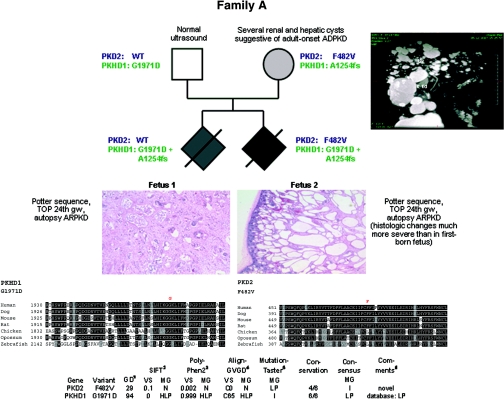
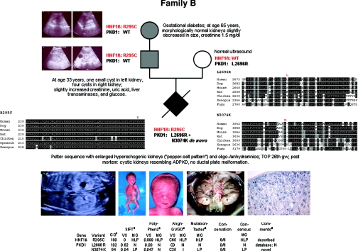
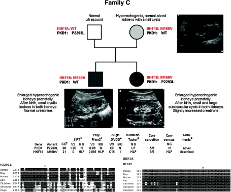
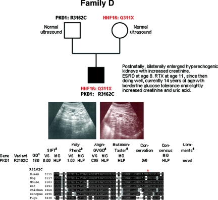
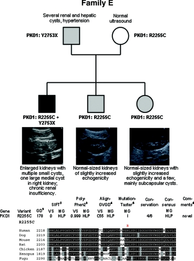
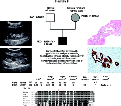
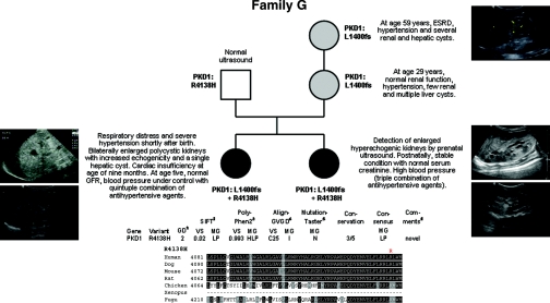
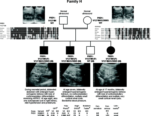
Autosomal dominant polycystic kidney disease (ADPKD) is typically a late-onset disease caused by mutations in PKD1 or PKD2, but about 2% of patients with ADPKD show an early and severe phenotype that can be clinically indistinguishable from autosomal recessive polycystic kidney disease (ARPKD). The high recurrence risk in pedigrees with early and severe PKD strongly suggests a common familial modifying background, but the mechanisms underlying the extensive phenotypic variability observed among affected family members remain unknown. Here, we describe severely affected patients with PKD who carry, in addition to their expected familial germ-line defect, additional mutations in PKD genes, including HNF-1β, which likely aggravate the phenotype. Our findings are consistent with a common pathogenesis and dosage theory for PKD and may propose a general concept for the modification of disease expression in other so-called monogenic disorders.


Figures

Figure 1.
Figure 1.
Pedigree of family A with clinical information, genotypes, multisequence alignments, and bioinformatic data for both missense changes detected. Renal histology shows much more severe changes in the second-born fetus than in the first-born as regards size and number of renal cysts. (Top right panel) MR cholangiogram (T2-weighted gadolinium-enhanced coronal projection) of the mother of both fetuses in family A at 35 years of age showing multiple cysts of various sizes. The multiple hepatic cysts do not communicate with the biliary tree. Liv, Liver; R Kd, right kidney; S, superior; I, inferior; LA, left anterior; RP, right posterior.
Figure 2.
Figure 2.
Pedigree of family B with genotypes, clinical and ultrasonographic data, multisequence alignments, and bioinformatic data for all three missense changes identified in this family. (Top panel) At age 65 years, the grandmother's kidneys were slightly decreased in size but morphologically normal without any cyst. (Middle panel) The father's kidneys showed one small cyst on the left and four cysts on the right side at the age of 33 years. (Bottom left panel) Prenatal ultrasound revealed oligo/anhydramnios and enlarged hyperechogenic fetal kidneys with a so-called “pepper-salt pattern.” (Bottom middle panel) Post mortem findings in the severely affected male fetus of this family with facial features typical of Potter sequence (termination of pregnancy in the 26th week of gestation [TOP 26th gw]). Abdominal situs with bilaterally enlarged kidneys (weight, 12.5 g; reference range for respective gestational age, 5.5 to 9.3 g). *Kidneys; +adrenal glands; arrows, ureters. (Bottom right panel) Renal histology (2.5× hematoxylin and eosin staining) with small tubular and glomerular cysts. The collecting ducts were not considerably dilated.
Figure 3.
Figure 3.
Pedigree of family C with genotypes, in silico information on both missense changes detected in this family, and clinical and ultrasonographic data. Mother and son presented with hyperechogenic normal-sized kidneys with small cysts, whereas the daughter additionally displayed some large subcapsular cystic lesions.
Figure 4.
Figure 4.
Pedigree and genotypes of family D. Renal ultrasound of the propositus at the age of 10 years with polycystic, considerably enlarged hyperechogenic kidneys. (Bottom panel) Bioinformatic data obtained for the PKD1 mutation R3162C.
Figure 5.
Figure 5.
Pedigree, genotypes, and renal ultrasound data of all children of family E. Enlarged kidneys with multiple small cysts and one large medial cyst in the right kidney were present in the eldest son, whereas his younger siblings demonstrated normal-sized kidneys with slightly increased echogenicity. A few mainly subcapsular cysts were also seen in the girl. (Bottom panel) In silico data obtained for the PKD1 mutation R2255C identified in this pedigree.
Figure 6.
Figure 6.
Pedigree and genotypes of family F. (Left panel) Renal ultrasound of the propositus at the age of 15 years with increased echogenicity, one large cyst in the left kidney (bottom panel) and loss of corticomedullary differentiation. (Right top panel) Liver histology at the age of 9 years showing ductal plate malformation with irregularly distributed, dilated portal vein branches (asterisk), and dilated bile ducts (cross) in a fibrotic, expanded portal field (hematoxylin and eosin; original magnification, ×40). (Right bottom panel) Liver histology at the age of 15 years in line with ductal plate malformation and congenital hepatic fibrosis and demonstrating a portal tract with irregular, circular arrangement of widened bile ducts (brown) that extend into the hepatic lobules. Note absence of inflammation and fibrosis in this portal tract (Cytokeratin 7 immunoperoxidase staining; original magnification, ×200). (Bottom panel) Bioinformatic prediction scores and multiple sequence alignments obtained for the PKD1 change L2696R.
Figure 7.
Figure 7.
Pedigree and genotypes of family G. (Left panel) Renal ultrasound of proposita postnatally (left top panel) and at the age of 4 years (left bottom panel). (Right top panel) Renal ultrasound of maternal grandmother at the age of 59 years. (Middle and bottom right panels) Renal ultrasound of the second-born daughter postnatally (middle right panel) and at the age of 15 months (bottom right panel). (Bottom panel) Bioinformatic predictions and multiple sequence alignments for the PKD1 variant R4138H identified on the paternal allele of both severely affected girls.
Figure 8.
Figure 8.
Pedigree, ultrasonographic data, and genotypes of consanguineous family H. (Top left and right panels) Normal renal ultrasound data of the parents at age 25 (mother) and 29 (father). (Bottom panel) Renal ultrasonographic data of all three affected children of this family with enlarged polycystic kidneys with loss of corticomedullary differentiation and bioinformatic information on both missense changes identified in this family.

References

    1. Wilson PD: Polycystic kidney disease. N Engl J Med 350: 151–164, 2004 - PubMed
    1. Harris PC, Torres VE: Polycystic kidney disease. Annu Rev Med 60: 321–337, 2009 - PMC - PubMed
    1. Torres VE, Harris PC, Pirson Y: Autosomal dominant polycystic kidney disease. Lancet 369: 1287–1301, 2007 - PubMed
    1. Bergmann C, Zerres K: Early manifestations of polycystic kidney disease. Lancet 369: 2157, 2007 - PubMed
    1. Ogborn MR: Polycystic kidney disease: A truly pediatric problem. Pediatr Nephrol 8: 762–767, 1994 - PubMed

Publication types

MeSH terms

Substances

LinkOut - more resources