. 2011 Dec 21;22(12):2383-9.
doi: 10.1021/bc200405d. Epub 2011 Nov 7.

89Zr-labeled dextran nanoparticles allow in vivo macrophage imaging

Affiliations

89Zr-labeled dextran nanoparticles allow in vivo macrophage imaging

Edmund J Keliher et al. Bioconjug Chem. .

Abstract

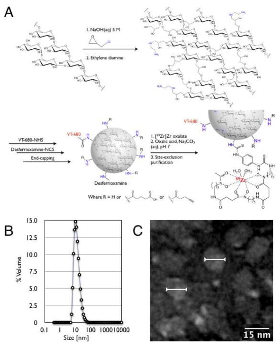
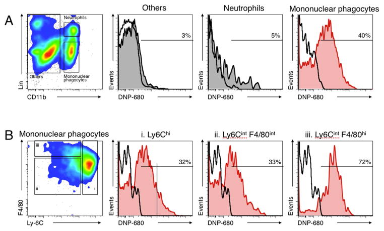
Tissue macrophages play a critical role both in normal physiology and in disease states. However, because of a lack of specific imaging agents, we continue to have a poor understanding of their absolute numbers, flux rates, and functional states in different tissues. Here, we describe a new macrophage specific positron emission tomography imaging agent, labeled with zirconium-89 ((89)Zr), that was based on a cross-linked, short chain dextran nanoparticle (13 nm). Following systemic administration, the particle demonstrated a vascular half-life of 3.9 h and was found to be located primarily in tissue resident macrophages rather than other white blood cells. Subsequent imaging of the probe using a xenograft mouse model of cancer allowed for quantitation of tumor-associated macrophage numbers, which are of major interest in emerging molecular targeting strategies. It is likely that the material described, which allows the visualization of macrophage biology in vivo, will likewise be useful for a multitude of human applications.


Figures

Figure 1
Figure 1
(A) General synthesis and radiolabeling of dextran nanoparticles (DNPs). (B) Dynamic laser light scattering data for the 13 nm DNP preparation. C. Transmission electron microscopy image of several individual DNPs measuring 13 nm in diameter on average (arrows).
Figure 2
Figure 2
Pharmacokinetics of different DNP. (A) Effect of size on blood half-life. (B) Effect of surface modification on blood half-life. (C) Biodistribution plotted as tissue concentration 24 h after intravenous administration of succinylated 13 nm DNP. Insert: Distribution to major organs.
Figure 3
Figure 3
PET imaging of 89Zr-DNP in mice bearing bilateral flank tumors (24 h after administration). (A and B) two different animals showing similar distribution to all 4 tumors (coronal stacks). (C) Three-dimensional rendering of animal presented in A.
Figure 4
Figure 4
Autoradiography and histology of DNP distribution. (A) Autoradiography of 1 mm tumor and other tissue sections show predominant accumulation in tumors. (B) Co-localization of representative autoradiography sections with adjacent Mac3 immunohistology sections. (C) Distribution of fluorescent DNP to Mac3 positive cells.
Figure 5
Figure 5
Microscopy of DNP distribution in tumor microenvironment in live mouse. (A) 3 h after injection; (B) 24 h after injection. Initially the DNP is primarily seen in vasculature whereas it is almost exclusively co-localized (yellow color) with TAM at 24 h.
Figure 6
Figure 6
(A) Flow cytometric identification of mononuclear phagocytes (CD11b+ Linlow), neutrophils (CD11b+ Linhigh (Ly-6G+)), and other cells (CD11b) in the tumor stroma (left). The three histograms on the top right show the percent of each population with detectable DNP-680 particles. Nearly all fluorescent DNP (DNP-680) is associated with mononuclear phagocytes; (B) Flow cytometric identification of mononuclear phagocyte subsets. CD11b+ Linlow cells were further divided into Ly6Chi (i) and Ly6Cint F4/80int (ii) monocyte-like cells and Ly-6Cint F4/80hi macrophages (left). The three histograms on the right show the percent of each population with detectable DNP-680 particles. DNP is associated with both monocyte subsets (Ly6Chi and Ly6Clo) as well as with macrophages (F4/80hi).

References

    1. Lee SH, Starkey PM, Gordon S. Quantitative analysis of total macrophage content in adult mouse tissues. Immunochemical studies with monoclonal antibody F4/80. J Exp Med. 1985;161:475–489. - PMC - PubMed
    1. Ho MK, Springer TA. Tissue distribution, structural characterization, and biosynthesis of Mac-3, a macrophage surface glycoprotein exhibiting molecular weight heterogeneity. J Biol Chem. 1983;258:636–642. - PubMed
    1. Di Gregorio GB, Yao-Borengasser A, Rasouli N, Varma V, Lu T, Miles LM, Ranganathan G, Peterson CA, McGehee RE, Kern PA. Expression of CD68 and macrophage chemoattractant protein-1 genes in human adipose and muscle tissues: association with cytokine expression, insulin resistance, and reduction by pioglitazone. Diabetes. 2005;54:2305–2313. - PubMed
    1. Thiele J, Braeckel C, Wagner S, Falini B, Dienemann D, Stein H, Fischer R. Macrophages in normal human bone marrow and in chronic myeloproliferative disorders: an immunohistochemical and morphometric study by a new monoclonal antibody (PG-M1) on trephine biopsies. Virchows Arch A Pathol Anat Histopathol. 1992;421:33–39. - PubMed
    1. Gaglia JL, Guimaraes AR, Harisinghani M, Turvey SE, Jackson R, Benoist C, Mathis D, Weissleder R. Noninvasive imaging of pancreatic islet inflammation in type 1A diabetes patients. J Clin Invest. 2011;121:442–445. - PMC - PubMed

Publication types