. 2011 Dec;179(6):2931-40.
doi: 10.1016/j.ajpath.2011.08.043. Epub 2011 Oct 28.

A novel strategy to develop therapeutic approaches to prevent proliferative vitreoretinopathy

Affiliations

A novel strategy to develop therapeutic approaches to prevent proliferative vitreoretinopathy

Steven Pennock et al. Am J Pathol. 2011 Dec.

Abstract

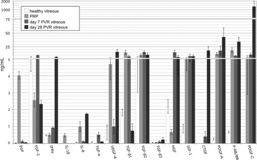
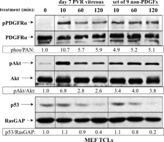
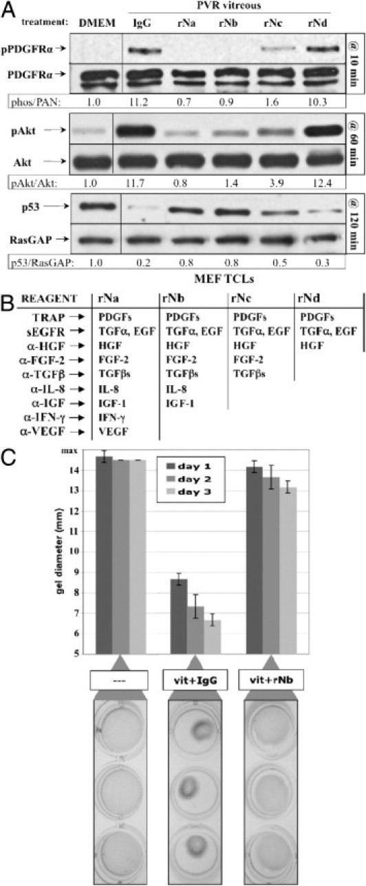
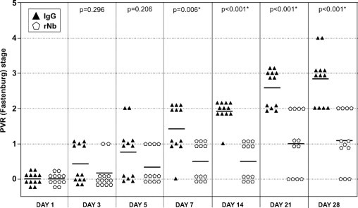
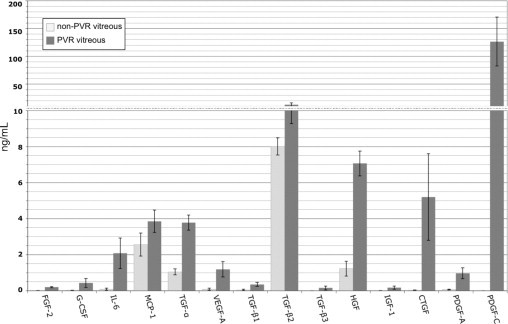
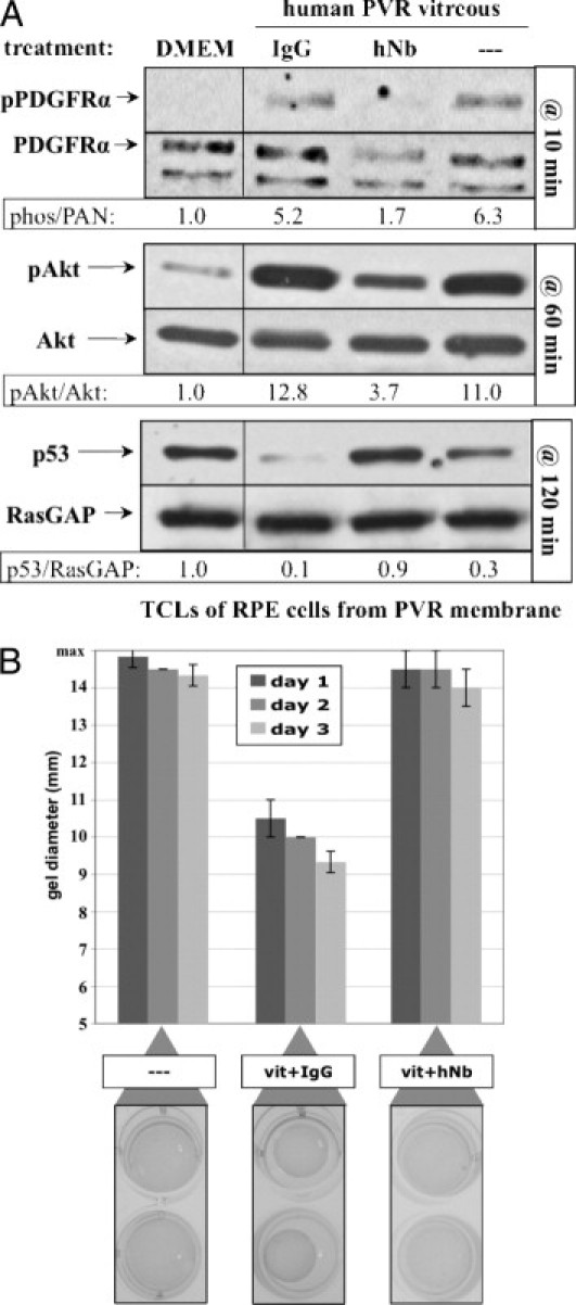
Proliferative vitreoretinopathy (PVR) thwarts the repair of rhegmatogenous retinal detachments. Currently, there is no effective prevention for PVR. Platelet-derived growth factor receptor α (PDGFRα) is associated with PVR in humans and strongly promotes experimental PVR driven by multiple vitreal growth factors outside the PDGF family. We sought to identify vitreal factors required for experimental PVR and to establish a potential approach to prevent PVR. Vitreous was obtained from normal rabbits or those in which PVR was either developing or stabilized. Normal vitreous contained substantial levels of growth factors and cytokines, which changed quantitatively and/or qualitatively as PVR progressed and stabilized. Neutralizing a subset of these agents in rabbit vitreous eliminated their ability to induce PVR-relevant signaling and cellular responses. A single intravitreal injection of neutralizing reagents for this subset prevented experimental PVR. To identify growth factors and cytokines likely driving PVR in humans, we subjected vitreous from patients with or without PVR to a similar series of analyses. This analysis accurately identified those agents required for vitreous-induced contraction of cells from a patient PVR membrane. We conclude that combination therapy encompassing a subset of vitreal growth factors and cytokines is a potential approach to prevent PVR.


Figures

Figure 1
Figure 1
The level of growth factors and cytokines in rabbit vitreous with or without PVR. Vitreous or platelet-rich plasma (PRP) was isolated from rabbits and subjected to either multiplex or Western blot analysis to quantify the level of 24 growth factors or cytokines. Healthy vitreous indicates vitreous isolated from uninjected eyes; PVR vitreous designates vitreous isolated from rabbits that were subjected to the PVR protocol and developed a PVR score of stage 3 or higher. The levels of G-CSF, IL-6, IL-10, MCP-1, TNF-α, TNF-β, and GM-CSF were all below detection (ie, <0.1 ng/mL) and not shown in the graph. Only the active forms of the three TGF-β isoforms were quantified. The bars represent the mean, whereas error bars indicate SD. Vitreous from 10 to 11 rabbits was used in this analysis; within each group, 5 or 6 were used for multiplex analysis and the remainder for Western blot analysis. PRP from three rabbits was used for multiplex analysis and from a second group of three rabbits for Western blot analysis. Three scales, contiguously aligned, were used along the y axis to span the entire range of concentrations measured.
Figure 2
Figure 2
Signaling events induced by PVR vitreous or the set of 9 non-PDGFs. Serum-starved MEFs were treated for the indicated times with either day 7 rabbit PVR vitreous, or the set of 9 non-PDGFs. Each agent in this set was used at the concentration measured in day 7 rabbit vitreous: FGF-2 (9.5 ng/mL), IGF-1 (8.3 ng/mL), TGF-β1 (7.7 ng/mL), TFG-α (0.5 ng/mL), EGF (0.12 ng/mL), IFN-γ (1.0 ng/mL), IL-8 (0.12 ng/mL), VEGF (1.0 ng/mL), and HGF (15.0 ng/mL). Cells were lysed and subjected to Western blot analysis using the indicated antibodies. Ratios representing normalized band intensities are shown under each pair of immunoblots. The data shown are representative of three independent experiments. TCL, total cell lysate.
Figure 3
Figure 3
Neutralizing growth factors and cytokines inhibited PVR vitreous–induced signaling events and gel contraction. A: rNb prevented PVR vitreous–induced signaling. Serum-starved MEFs were treated for the indicated duration at 37°C with DMEM alone, day 7 PVR vitreous supplemented with either isotype-control IgG, or one of the four neutralization mixtures (rNa, rNb, rNc, or rNd) listed in B. Cells were harvested, and the resulting samples processed as described in Figure 2. All immunoblots shown are representative of three independent experiments. TCL, total cell lysate. B: Table of neutralization components (left column) and their respective growth factor/cytokine targets (right columns). C: rNb prevented PVR vitreous–induced contraction of collagen gels. MEFs were subjected to the collagen gel contraction assay (Materials and Methods) in the presence of either DMEM (—), or day 7 PVR vitreous (vit) supplemented with IgG or rNb. Gel diameter was measured manually, and data presented are a mean of three independent experiments ± SD (shown as error bars). Images of triplicate wells from a single representative experiment at day 2 are shown below the graph.
Figure 4
Figure 4
Neutralizing a subset of vitreal growth factors and cytokines prevented retinal detachment in rabbits. One week after a gas vitrectomy, rabbits received three separate 0.1-mL injections of primary rabbit conjunctival fibroblasts, platelet-rich plasma, and either a 50-fold molecular excess of rNb neutralization mixture (12 rabbits, pentagons), or the equivalent amount of isotype control IgG (12 rabbits, triangles). Rabbits were examined and scored for PVR at the indicated times. Horizontal bars represent the mean of each group. Statistically significant differences between the two groups at each time point were determined by Mann-Whitney analysis (P values are indicated above each time point). Fundus photographs were not recorded because the morphology for a given stage was essentially the same as previously reported.
Figure 5
Figure 5
The level of growth factors and cytokines in vitreous of patients with or without PVR. Same as Figure 1 except that the analysis was performed on vitreous from patient donors. Non-PVR ocular pathologies included macular holes or macular puckers. Both the non-PVR and PVR groups consisted of 12 patients; data represent the mean values for each group, whereas error bars show SD. The levels of EGF, IFN-γ, IL-1β, IL-10, TNF-α, TNF-β, GM-CSF, and PDGF-AB/BB were all below detection (ie, <0.1 ng/mL) in all samples and thus omitted from the graph. Only the active forms of the three TGF-β isoforms were quantified. Statistically significant differences (P < 0.05) existed between non-PVR and PVR vitreal levels for the following growth factors/cytokines: FGF-2, G-CSF, IL-6, TGF-α, VEGF, CTGF, TGF-βs, HGF, IGF-1, and PDGFs. Two scales were used along the y axis to adequately span the entire range of concentrations.
Figure 6
Figure 6
A neutralizing cocktail inhibited human PVR vitreous–induced signaling events and gel contraction. A: Neutralization of a subset of vitreal growth factors and cytokines prevented PVR vitreous–induced signaling. Subconfluent cultures of human RPE cells isolated from a PVR membrane were serum starved overnight and then treated for the indicated time at 37°C with either DMEM, or human PVR vitreous supplemented with nothing (—), nonimmune IgG, or hNb (composition described in Supplemental Figure S2B available at http://ajp.amjpathol.org). After stimulation, cells were lysed, and the resulting samples were processed as described in Figure 2. All immunoblots shown are representative of three independent experiments. TCL, total cell lysate. B: hNb prevented PVR vitreous–induced contraction of collagen gels. Human RPE cells isolated from a PVR membrane were subjected to the collagen gel contraction assay (Materials and Methods) in the presence of either DMEM (—), or vitreous (vit) from a PVR patient donor supplemented with IgG or hNb. Gel diameter was measured manually and data presented are a mean of three independent experiments ± SD (shown as error bars). Images of duplicate wells from a single representative experiment at day 2 are shown below the graph. Human vitreous used in this experiment was taken from the same pool used in Supplemental Figure S2 (available at http://ajp.amjpathol.org).

References

    1. Han D. In: Albert D., JW M., DT A., BA B., editors. Elsevier Saunders; Philadelphia, PA: 2008. Proliferative vitreoretinopathy; pp. 2315–2324.
    1. Pastor J.C. Proliferative vitreoretinopathy: an overview. Surv Ophthalmol. 1998;43:3–18. - PubMed
    1. Michels R.G., Wilkinson C.P., Rice A. Mosby; St. Louis, MO: 1990. Proliferative retinopathy.
    1. Glaser B.M., Cardin A., Biscoe B. Proliferative vitreoretinopathy: The mechanism of development of vitreoretinal traction. Ophthalmology. 1987;94:327–332. - PubMed
    1. Campochiaro P.A. Mechanisms in ophthalmic disease: pathogenic mechanisms in proliferative vitreoretinopathy. Arch Ophthalmol. 1997;115:237–241. - PubMed

Publication types

MeSH terms

Substances