. 2011 Oct 28;147(3):539-53.
doi: 10.1016/j.cell.2011.10.003.

Endothelial-derived angiocrine signals induce and sustain regenerative lung alveolarization

Affiliations

Endothelial-derived angiocrine signals induce and sustain regenerative lung alveolarization

Bi-Sen Ding et al. Cell. .

Abstract

To identify pathways involved in adult lung regeneration, we employ a unilateral pneumonectomy (PNX) model that promotes regenerative alveolarization in the remaining intact lung. We show that PNX stimulates pulmonary capillary endothelial cells (PCECs) to produce angiocrine growth factors that induce proliferation of epithelial progenitor cells supporting alveologenesis. Endothelial cells trigger expansion of cocultured epithelial cells, forming three-dimensional angiospheres reminiscent of alveolar-capillary sacs. After PNX, endothelial-specific inducible genetic ablation of Vegfr2 and Fgfr1 in mice inhibits production of MMP14, impairing alveolarization. MMP14 promotes expansion of epithelial progenitor cells by unmasking cryptic EGF-like ectodomains that activate the EGF receptor (EGFR). Consistent with this, neutralization of MMP14 impairs EGFR-mediated alveolar regeneration, whereas administration of EGF or intravascular transplantation of MMP14(+) PCECs into pneumonectomized Vegfr2/Fgfr1-deficient mice restores alveologenesis and lung inspiratory volume and compliance function. VEGFR2 and FGFR1 activation in PCECs therefore increases MMP14-dependent bioavailability of EGFR ligands to initiate and sustain alveologenesis.


Figures

Figure 1
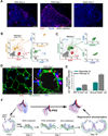
Figure 1. Unilateral left lung pneumonectomy (PNX) induces right lung regeneration and expansion of lung epithelial progenitors within 3 days post-PNX
(A, B) The kinetics of restoration of lung weight and volume in the remaining intact right lung lobes after resection of left lung. A, schema illustrating PNX procedure and representative image of the remaining regenerated right lungs 15 days after PNX. B. Lung regeneration is initiated 3 days after PNX and achieves its maximum size and volume at day 15 after PNX. n = 5. Data are presented as mean ± s.e.m, throughout. (C) Amplification of CCSP+ cells at bronchioalveolar duct junction (BADJ) on day 3 after PNX. Mice were fed with BrdU-containing drinking water to pulse proliferating lung progenitors. There is a specific expansion of CCSP+BrdU +cells localized at BADJ on day 3 after PNX (arrows). Note the distribution of BrdU+ cells in distal alveolar space thereafter (arrowheads). (D & E) CCSP+SPC+Sca-1+VE-cadherinCD31 BASC-like cells were identified and quantified in CCSP-YFP and SPC-YFP mice 3 days after PNX. There is minimal BrdU uptake in the VE-cadherin+CD31+ PCECs, indicating that at this time point PCECs do not undergo proliferation. Note the close cellular juxtaposition of VE-cadherin+ PCECs (blue arrow) and proliferating CCSP+BrdU+ BASCs (red arrow) in lower inset of D panel.
Figure 2
Figure 2. At day 7 after PNX, expansion of AECs and PCECs sustain alveolar regeneration
(A) After PNX, proliferating cells were pulsed with intraperitoneal administration of BrdU and stained for BrdU at different time points. BrdU+ transit amplifying cells (TACs) increased significantly throughout the right lung and peaked at day 7 after PNX. Scale bar, 2.5 mm. (B, C) Quantification of TACs in the remaining right lungs at day 7 after PNX. Polyvariate flow cytometric analysis of total mononuclear lung cells 7 days after PNX demonstrating expansion of SPD+SPC+E-cadherin+ type II-like AECs (AECIIs) and VE-cadherin+CD34+VEGFR2+FGFR1+CD45 PCECs. (D) Proliferation of SPC+ AECIIs and VE-cadherin+ PCECs at the alveolar-capillary interface in the remaining lungs at day 7 after PNX. Note the close cellular proximity between PCECs (green arrow) with BrdU+ AECIIs (red arrow). Scale bar, 100 µm. (E) Quantification of VE-cadherin+CD34+ PCECs and SPC+E-cadherin+ AECIIs in the remaining right lungs 15 days after PNX; There is significant proliferation of both PCECs and AECs induced by PNX. n =5. (F) Proposed model for regenerative alveolarization mediated by proliferation of lung epithelial progenitors. PNX-induced regeneration is primarily mediated by amplification of BASCs, AECs and PCECs during alveolar regeneration.
Figure 3
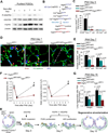
Figure 3. Inducible deletion of Vegfr2 and partial knockdown of Fgfr1 in endothelial cells (EC) attenuates lung regeneration
(A) Sequential activation of VEGFR2 and upregulation of FGFR1 in PCECs after PNX. VEGFR2 phosphorylation is increased by PNX, while the total VEGFR2 expression in PCECs remains constant. In contrast, FGFR1 expression in PCECs is significantly upregulated after PNX in a time-dependent manner. (B) Generation of EC-specific knockout of VEGFR2 and FGFR1 in adult mice. Transgenic mice in which VE-cadherin promoter drives the expression of tamoxifen-responsive CreERT2 (VE-Cad-CreERT2 mice) were crossed with Vegfr2loxP/loxP and Fgfr1loxP/loxP mice and treated with tamoxifen to induce EC-specific deletion of Vegfr2 and Fgfr1 (Vegfr2iΔEC/iΔEC and Vegfr2iΔEC/iΔECFgfr1iΔEC/+ mice). (C) EC-specific deletion of Vegfr2 (Vegfr2iΔEC/iΔEC mice) inhibits the expansion of CCSP+Sca1+ BASC-like cells after PNX. Vegfr2iΔEC/+ mice were used as control. (D, E) Defective proliferation of both PCECs (red arrowheads) and AECs (yellow arrows) in Vegfr2iΔEC/iΔECFgfr1iΔEC/+ mice after PNX, n = 4. Scale bar, 100 µm. Note that the increase in alveolar diameter (dashed arrows) in the Vegfr2iΔEC/iΔEC Fgfr1iΔEC/+ mice as compared to the control Vegfr2iΔEC/+ mice. (F) After PNX, EC-specific deletion of Vegfr2 and Fgfr1 impaired the recovery of pulmonary function. The restoration of the pulmonary function in the Vegfr2iΔEC/iΔECFgfr1iΔEC/+ mice was significantly inhibited compared to the control mice. Note the normal pulmonary function of knockout mice at steady state conditions prior to PNX. #, p < 0.01, compared to control Vegfr2iΔEC/+ mice, n = 4. (G) Restoration of lung mass and volume is impaired in mice with EC-specific deletion of Vegfr2 and Fgfr1, n = 4. (H) Proposed model of PCEC-mediated regulation of regenerative alveolarization. Activation of VEGFR2 in PCECs instigates the early expansion of BASCs, while there is minimal proliferation of PCECs. Subsequent upregulation of FGFR1 along with VEGFR2 activation sustains proliferation of PCECs and AECs that peaks at day 7. PCECs through neo-vascularization and inducing AEC expansion complete regeneration of the right lungs by day 15 after PNX.
Figure 4
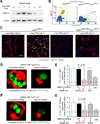
Figure 4. After PNX, MMP14 is specifically produced by PCECs and induces formation of alveolar-capillary-like sacs in 3-dimensional coculture with activated ECs
(A) PNX induces a time-dependent upregulation of MMP14 protein in the remaining lobe of the lung.Representative Western blot image is shown. (B, C) After PNX, specific upregulation of MMP14 in VE-cadherin+ PCECs is attenuated in Vegfr2iΔEC/iΔECFgfr1iΔEC/+ mice, as shown by flow cytometry (B) and immunostaining (C). Note the colocalization of upregulated MMP14 in VE-cadherin+ PCECs (arrow), but not ECs in the liver of pneumonectomized control mice. Scale bar, 100 µm. (D, E) Formation of 3-dimensional spheroids with MAP kinase activated ECs (MAPK-ECs) establishes a bioreactor for the expansion of SPC (YFP)+ AECs, which is dependent on the angiocrine production of MMP14. Representative image (D) and quantification (E) of different groups are shown. scr, scrambled shRNA, CM, conditioned medium. (F, G) Angiocrine production of MMP14 supports the propagation of CCSP (YFP)+Sca-1+CD31BASC-like cells. Representative image (F) and quantification (G) of various groups are shown.
Figure 5
Figure 5. PCEC-derived MMP14 supports regenerative alveolarization
(A) After PNX, neutralizing mAb to MMP14 abolished the regeneration of lung mass and volume. (B) After PNX, inhibition of MMP14 diminished expansion of the E-cadherin+ AECs, n = 5. Scale bar, 100 µm. Note the lack of both cuboidal SPC+E-cadherin+ AECIIs (yellow arrow) and squamous SPCE-cadherin+ type I-like AECs (red arrowhead) in the alveoli treated with MMP14 mAb. (C, D) After PNX, MMP14 inhibition primarily blocked the expansion of E-cadherin+ AECs, but not VE-cadherin+CD34+ PCECs, n=5. (E, F) After PNX, inhibition of MMP14 suppressed alveolar regrowth and led to enlarged alveolar size. E) Representative H&E staining of the pneumonectomized lungs treated with neutralizing mAb to MMP14 and isotype IgG. Note the increase in the alveolar size in the mAb treated mice (dashed lines). F) Quantification of alveolar number and alveolar size after PNX. Scale bar, 100 um.
Figure 6
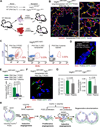
Figure 6. Angiocrine production of MMP14 induces alveologenesis by shedding EGF-like ectodomains from HB-EGF and laminin5 γ2 chain, activating EGF receptor (EGFR)
(A, B) PNX induced time-dependent release of HB-EGF into alveolar space, which is inhibited in Vegfr2iΔEC/iΔECFgfr1iΔEC/+ mice or by MMP14 neutralization. Representative Western blot image is shown. Control Vegfr2iΔEC/+ mice treated with neutralizing mAb to MMP14 (MMP14 Ab); BAL,bronchioalveolar lavage. BALF, BALF fluid; n = 4. (C) At day 7 after PNX, activation of VEGFR2 and FGFR1 in PCECs and production of MMP14 caused cleavage of laminin5 γ2 chain. Representative Western blot image is shown. (D–F) EGF injection restored 1) regeneration of lung mass and volume (D), 2) integration of E-cadherin+AECs within the capillary (E) and 3) pulmonary function measured by inspiratory volume and static compliance (F) in Vegfr2iΔEC/iΔECFgfr1iΔEC/+ mice after PNX. Note the enhanced association of SPCE-cadherin+ AECIIs (red arrowhead) and SPC+E-cadherin+ AECIIs (yellow arrow) with the capillary. (G–I) At day 7 after PNX, intravenous EGF injection restored EGFR phosphorylation (G) and increased proliferation of SPC+ AECIIs (H, I) in the lungs of Vegfr2iΔEC/iΔECFgfr1iΔEC/+ mice. Note the augmented proliferation in SPC+ AECIIs (white arrow). Quantification of amplifying cell population after PNX is shown in I, n = 4. Scale bar, 100 µm.
Figure 7
Figure 7. Transplantation of wild type PCECs restores the defective alveolar regeneration in the mice deficient in endothelial Vegfr2 and Fgfr1
(A) Endothelial cell (EC) transplantation strategy to define the contribution of PCECs in promoting alveolar regeneration. After PNX, ECs were purified from the lung and liver of wild type (WT) littermates, transduced with lentiviral GFP, and transplanted through the jugular vein into Vegfr2iΔEC/iΔEC mice at day 3 after PNX and Vegfr2iΔEC/iΔECFgfr1iΔEC/+ mice at day 7 after PNX. (B) Incorporation of transplanted GFP+ PCECs into functional lung capillary. Intravenous infusion of vascular-specific isolectin from griffonia simplicifolia (GS-IB4) was used to identify functional vasculature. Note the presence of perfused isolectin+GFP+ PCECs indicating functional incorporation of transplanted WT PCECs into recipient Vegfr2iΔEC/iΔECFgfr1iΔEC/+capillaries. Scale bar, 100 µ m. (C & D) Restoration of the expansion potential of CCSP+ BASC-like cells in Vegfr2iΔEC/iΔEC mice after PCEC transplantation. Note in (D) the unique localization of proliferating BrdU+CCSP+ BASC-like cells (red arrow) that is in close adjacency to the transplanted GFP+ PCECs (green arrow). (E, F & G) Transplantation of WT PCECs restores proliferation of SPC+ AECs (E, F) and pulmonary function (G) in Vegfr2iΔEC/iΔECFgfr1iΔEC+ mice. Expanding BrdU+SPC+ AECs (red arrow) were detected in close cellular association with transplanted PCECs (green arrow) (F). (H) Proposed model illustrating the inductive role of VEGFR2 and FGFR1 primed PCECs in lung regenerative alveolarization. Upon PNX, activation of VEGFR2 in PCECs leads to MMP14 production and HB-EGF release to stimulate the expansion of epithelial progenitor cells (BASCs and AECIIs). Subsequent activation of FGFR1 along with VEGFR2 stimulates proliferation of PCECs maintaining MMP14 expression. MMP14 unmasks cryptic EGFR ligands through shedding of HB-EGF and cleaving laminin5 γ2 chain, which by activating EGFR induce proliferation of SPC+E-cadherin+ AECs. After PNX, sequential propagation of epithelial cells induced by PCEC-derived MMP14 and increase in bioavailability of EGFR ligands, culminates in full reconstitution of physiologically functional alveolar-capillary sacs. Proliferation of the PCECs mediated through VEGFR2 and FGFR1, vascularizes the regenerating lung tissue to restore the blood supply and gas exchange function.

Comment in

References

    1. Aird WC. Phenotypic heterogeneity of the endothelium: I. Structure, function, and mechanisms. Circ Res. 2007;100:158–173. - PubMed
    1. Alvarez DF, Huang L, King JA, ElZarrad MK, Yoder MC, Stevens T. Lung microvascular endothelium is enriched with progenitor cells that exhibit vasculogenic capacity. Am J Physiol Lung Cell Mol Physiol. 2008;294:L419–430. - PubMed
    1. Atkinson JJ, Holmbeck K, Yamada S, Birkedal-Hansen H, Parks WC, Senior RM. Membrane-type 1 matrix metalloproteinase is required for normal alveolar development. Dev Dyn. 2005;232:1079–1090. - PubMed
    1. Beers MF, Kim CY, Dodia C, Fisher AB. Localization, synthesis, and processing of surfactant protein SP-C in rat lung analyzed by epitope-specific antipeptide antibodies. J Biol Chem. 1994;269:20318–20328. - PubMed
    1. Beers MF, Morrisey EE. The three R's of lung health and disease: repair,remodeling, and regeneration. J Clin Invest. 2011;121:2065–2073. - PMC - PubMed

Publication types

MeSH terms

Substances