. 2011 Oct 28;147(3):554-64.
doi: 10.1016/j.cell.2011.09.035.

The Eph-receptor A7 is a soluble tumor suppressor for follicular lymphoma

Affiliations

The Eph-receptor A7 is a soluble tumor suppressor for follicular lymphoma

Elisa Oricchio et al. Cell. .

Abstract

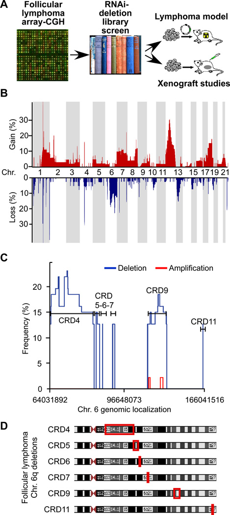
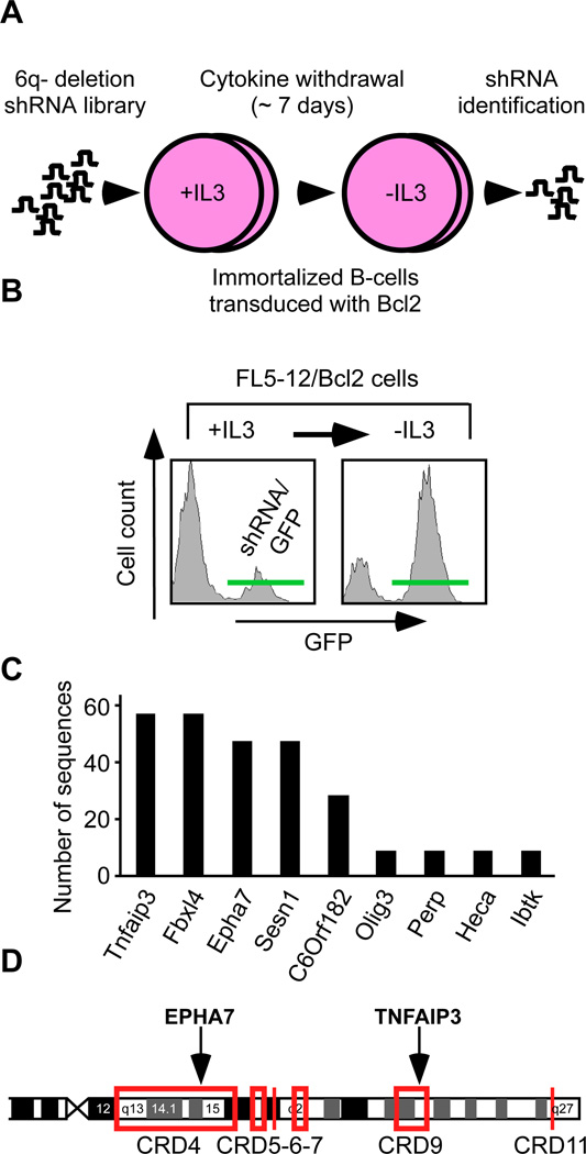
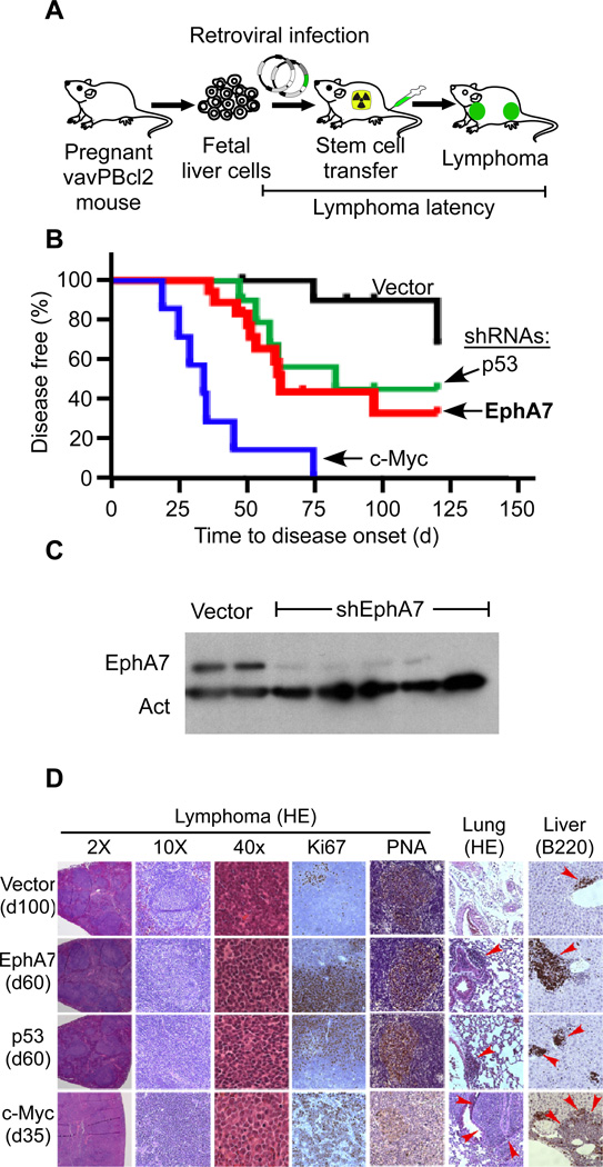
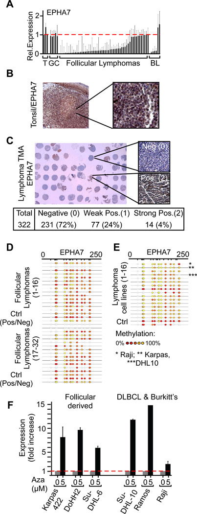
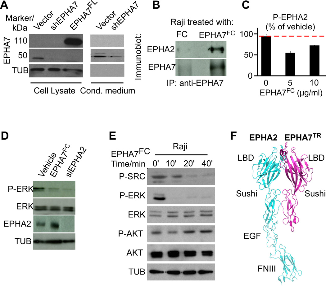
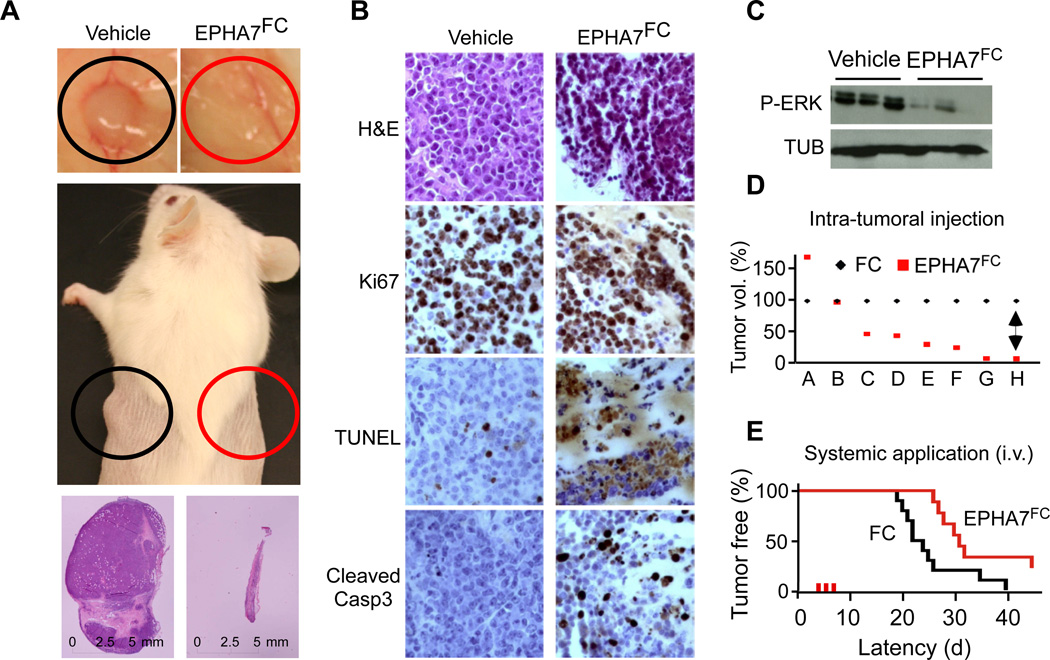
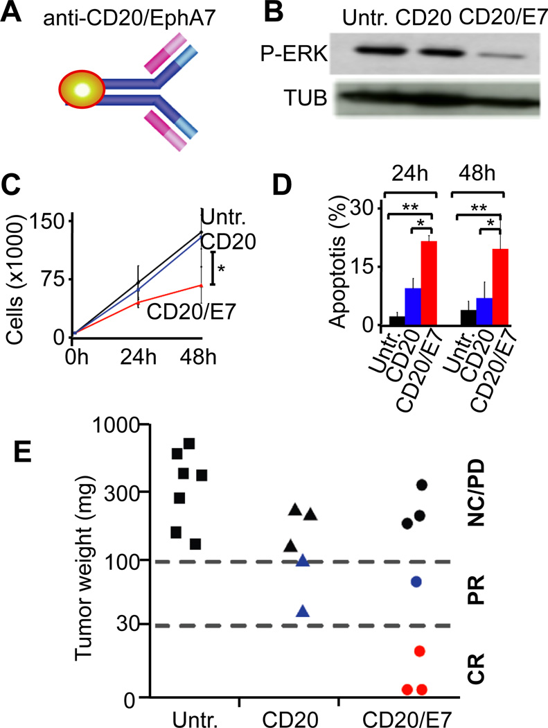
Insights into cancer genetics can lead to therapeutic opportunities. By cross-referencing chromosomal changes with an unbiased genetic screen we identify the ephrin receptor A7 (EPHA7) as a tumor suppressor in follicular lymphoma (FL). EPHA7 is a target of 6q deletions and inactivated in 72% of FLs. Knockdown of EPHA7 drives lymphoma development in a murine FL model. In analogy to its physiological function in brain development, a soluble splice variant of EPHA7 (EPHA7(TR)) interferes with another Eph-receptor and blocks oncogenic signals in lymphoma cells. Consistent with this drug-like activity, administration of the purified EPHA7(TR) protein produces antitumor effects against xenografted human lymphomas. Further, by fusing EPHA7(TR) to the anti-CD20 antibody (rituximab) we can directly target this tumor suppressor to lymphomas in vivo. Our study attests to the power of combining descriptive tumor genomics with functional screens and reveals EPHA7(TR) as tumor suppressor with immediate therapeutic potential.


Figures

Figure 1
Figure 1. Oncogenomic study to identify tumor suppressor genes in follicular lymphoma (FL)
A, The study design combines genomic tumor analyses with an RNAi screen and validation in murine models and in xenografts; B, Array-CGH analysis of 64 follicular lymphomas showing frequencies of genomic gain (red) and loss (blue) across the genome; C, High resolution depiction of recurrent gains (red) and losses (blue) affecting chromosome 6q, indicated are common regions of deletion (CRDs found in > 10%); D, Mapping of CRDs. The observed 6q deletions are typically large and hemizygous and do not readily identify a target gene. See also Figure S1.
Figure 2
Figure 2. A 6q-deletion specific RNAi screen functionally identifies EPHA7 as a candidate tumor suppressor gene
A, Design of a pooled, deletion-specific shRNA library screen in a surrogate model (immortalized FL5-12/Bcl2 cells); B, FACS profiles for GFP showing enrichment of cells expressing the shRNA library (and the GFP reporter) following IL-3 depletion. See also Figure S2; C, Absolute number and identity of shRNA sequences retrieved from the enriched population; D, EPHA7 and TNFAip3 map to the CRD4 and CRD9 in FL. See also Figure S1.
Figure 3
Figure 3. EphA7 opposes tumor development in a murine model of FL
A, A mosaic model of FL based on vavPBcl2 transgenic mice; B, Tumor latencies for animals receiving vavPBcl2 transgenic HSCs transduced with empty vector (black, n = 11), or shRNAs against EphA7 (red, n = 18) and p53 (green, n = 9) or over expressing c-Myc (blue, n = 7). C, Immunoblot on FACS purified vavPBcl2 lymphoma cells expressing vector or an shRNA against EphA7 and probed as indicated. D, Microscopic pathology and immunohistochemistry of vavPBcl2 lymphomas expressing the indicated constructs, red arrows indicate infiltrating tumor cells. See also Figure S3.
Figure 4
Figure 4. EPHA7 is differentially silenced in FLs and expressed in germinal center (GC) B-cells
A, qRT-PCR results for EPHA7 in purified B-cells from reactive tonsils (T), GC B-cells (GC), purified B-cells from follicular lymphomas (FL), and Burkitt’s lymphomas (BL) (mean +/− standard deviation; p (tumor vs. normal < 0.05 for FL and BL); B, Immunohistochemical detection of the EPHA7 protein in a normal tonsil; C, Representative section of tissue microarrays (TMA) representing 322 human FLs and stained for EPHA7; D and E, Mass-array analysis of EPHA7 promoter methylation in 32 follicular lymphomas (D), and 16 human lymphoma lines (E) and positive / negative controls (Ctrl); the color scale indicates the degree of methylation (Red: 0%; yellow: 100%); f, qRT-PCR of EPHA7 mRNA levels in human lymphoma cell lines treated with 5’aza-2’-deoxycytidine (Aza); for all cell lines: p(untr. vs. Aza.) < 0.01). See also Figure S4.
Figure 5
Figure 5. EPHA7FC binds to EPHA2 and blocks oncogenic signals in lymphoma cells
A, Lysates and conditioned media from FL5-12/Bcl2 cells expressing vector, an shRNA against EphA7 (shEphA7), or full length EphA7 (EphA7FL) probed with an antibody against EPHA7. B, Immunoprecipitation of lysates from Raji cells treated with EPHA7FC (FC-tagged ectodomain of EPHA7) or FC, IP with anti-EPHA7 and probed against EPHA7 and EPHA2; C, ELISA assay for EPHA2 phosphorylation on Raji cells treated with EPHA7FC or vehicle (FC); D, Immunoblot on lysates of Raji cells treated with vehicle, EPHA7FC (5µg) or an siRNA against EPHA2; E, Immunoblot on Raji cells treated with 5µg/ml EPHA7FC for the indicated times; F, Model of the EPHA2 – EPHA7TR interaction based on the known structure of EPHA2 and its homology with EPHA7 (LBD, ligand binding domain, EGF, EGF-like domain, FNIII, fibronectin domain). See also Figure S5.
Figure 6
Figure 6. Exogenous administration of purified EPHA7 suppresses human lymphoma xenografts
A, Xenografted Raji lymphomas grown in the flank of NOD/SCID mice and treated three times on alternate days by intra-tumoral administration of 20 µg EPHA7FC (red circle) or vehicle (FC; black circle); B, Microscopic pathology on EPHA7FC treated and mock treated Raji lymphomas stained as indicated; C, Immunoblot on lysates of tumors treated with EPHA7FC or vehicle in vivo; D, Matched pair analysis of tumor volumes of eight (A–H) treated (red) and control (black) Raji lymphomas; E, Intravenous (i.v.) administration of vehicle (FC, black) or EPHA7FC (20µg, for 3 days, red) delays tumor development from 1×10⁶ injected Raji lymphoma cells. See also Figure S6.
Figure 7
Figure 7. Targeted delivery of EPHA7 to xenografted lymphomas using an anti-CD20-EPHA7 fusion antibody
A, Schematic of the anti-CD20/EPHA7 fusion antibody; B, Immunoblot on Raji cells that were untreated (Untr.), treated with anti-CD20 (CD20), or anti-CD20-EPHA7TR fusion (CD20/E7); C, Proliferation of Raji cells treated as indicated (*denotes p(CD20 vs. CD20/E7 < 0.05); D, Apoptosis of Raji cells 30 treated as indicated at 24h and 48h; * and ** denote significance (p < 0.05).; E, Mice bearing Raji xenografts (> 1cm3) left untreated (Untr.) or given 1µg of anti-CD20 (CD20) or anti-CD20-EPHA7TR (CD20/E7) for 5 days and collected 2 days after last treatment. Tumors (in matrigel) weighed ex vivo and classified as complete response (CR): 0 – 30 mg, partial responses (PR): 30–100 mg; no change (NC)/progressive disease (PD): > 100 mg. See also Figure S7.

Comment in

References

    1. Barr PM, Lazarus HM. Follicular non-Hodgkin lymphoma: long-term results of stem-cell transplantation. Curr Opin Oncol. 2008;20:502–508. - PubMed
    1. Bende RJ, Smit LA, van Noesel CJ. Molecular pathways in follicular lymphoma. Leukemia. 2007;21:18–29. - PubMed
    1. Bidere N, Ngo VN, Lee J, Collins C, Zheng L, Wan F, Davis RE, Lenz G, Anderson DE, Arnoult D, et al. Casein kinase 1alpha governs antigen-receptor-induced NF-kappaB activation and human lymphoma cell survival. Nature. 2009;458:92–96. - PMC - PubMed
    1. Calado DP, Zhang B, Srinivasan L, Sasaki Y, Seagal J, Unitt C, Rodig S, Kutok J, Tarakhovsky A, Schmidt-Supprian M, et al. Constitutive canonical NF-kappaB activation cooperates with disruption of BLIMP1 in the pathogenesis of activated B cell-like diffuse large cell lymphoma. Cancer Cell. 2010;18:580–589. - PMC - PubMed
    1. Chin L, Gray JW. Translating insights from the cancer genome into clinical practice. Nature. 2008;452:553–563. - PMC - PubMed

Publication types