. 2011 Oct 28;147(3):641-52.
doi: 10.1016/j.cell.2011.09.037.

Motor neuron position and topographic order imposed by β- and γ-catenin activities

Affiliations

Motor neuron position and topographic order imposed by β- and γ-catenin activities

Elena Y Demireva et al. Cell. .

Abstract

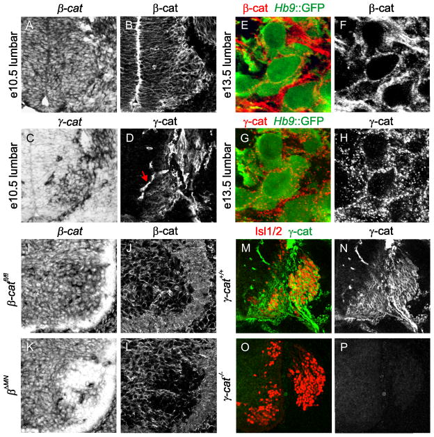
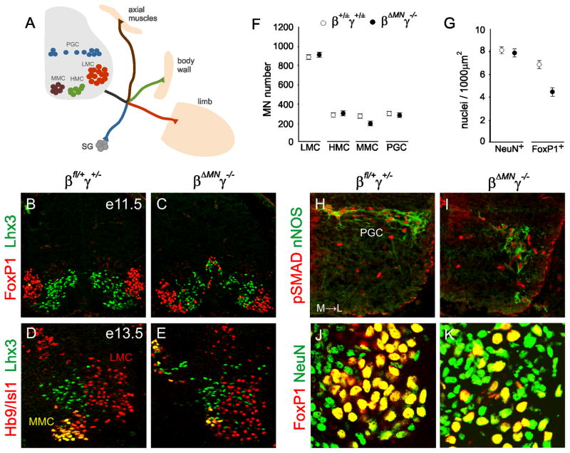
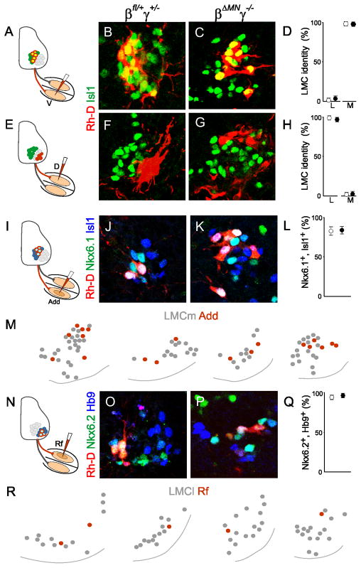
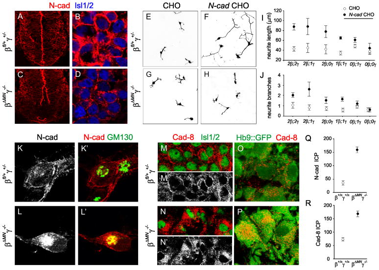
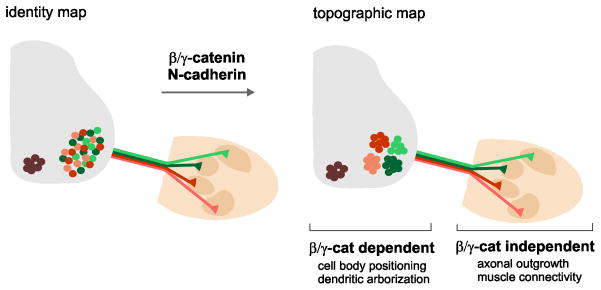
Neurons typically settle at positions that match the location of their synaptic targets, creating topographic maps. In the spinal cord, the organization of motor neurons into discrete clusters is linked to the location of their muscle targets, establishing a topographic map of punctate design. To define the significance of motor pool organization for neuromuscular map formation, we assessed the role of cadherin-catenin signaling in motor neuron positioning and limb muscle innervation. We find that joint inactivation of β- and γ-catenin scrambles motor neuron settling position in the spinal cord but fails to erode the predictive link between motor neuron transcriptional identity and muscle target. Inactivation of N-cadherin perturbs pool positioning in similar ways, albeit with reduced penetrance. These findings reveal that cadherin-catenin signaling directs motor pool patterning and imposes topographic order on an underlying identity-based neural map.


Figures

Figure 1
Figure 1. β- and γ-catenin expression in developing motor neurons
A,B. β-catenin (β-cat) expression in e10.5 lumbar spinal cord. C,D. γ-catenin (γ-cat) expression in e10.5 lumbar spinal cord. Arrow marks blood vessels. Between e15.5 and p0, γ-catenin transcript is extinguished from most spinal motor neurons (Figure S1E–H). E–H. β-cat and γ-cat expression in GFP+ lumbar motor neurons in e13.5 Hb9::GFP transgenic mice. I,K. β-cat expression in e13.5 lumbar spinal cord from control β-catfl/fl and βΔMN embryos. J,L. β-cat expression in e13.5 lumbar spinal cord from control β-catfl/fl and βΔMN embryos. In the absence of motor neuron β-catenin expression we observed a marked upregulation of γ-catenin protein (Figure S1I–L). M–P. Absence of γ-cat expression in e11.5 lumbar spinal cord of γ-cat−/− mice. RNA expression data in panels A, C, I and K with positive signal in black.
Figure 2
Figure 2. Motor neuron columnar segregation in β- and γ-catenin mutants
A. Topographic order in motor innervation of the mouse limb: median motor column (MMC) neurons innervate axial muscles, hypaxial motor column (HMC) neurons innervate body wall muscles, lateral motor column (LMC) neurons innervate limb muscles, and preganglionic motor column (PGC) neurons innervate sympathetic ganglion (SG) neurons. B–C. Segregation of Lhx3+ MMC and FoxP1+ LMC neurons at lumbar levels of e11.5 control and β ΔMNγ−/− embryos. D–E. Segregation of Lhx3+, Hb9+, Isl1+ MMC and Hb9+, Isl1+ LMC neurons at lumbar levels of e13.5 control and βΔMNγ−/− embryos. F. Post-mitotic motor neurons in the marginal zone of e13.5 spinal cord of control (β+/±γ+/±) and βΔMNγ−/− embryos. Motor neurons/100μm: mean +/− SEM (difference from control significant for MMC neurons; t-test, p<0.0001). Control catenin group (β+/±γ+/±) includes genotypes: β+/+γ+/+ (wt), β+/ΔMNγ+/+, β+/+γ+/− and β+/ΔMNγ+/−. G. NeuN+ and FoxP1+ neuronal densities in the LMC of e13.5 control and βΔMNγ−/− embryos; mean +/− SEM (t-test, p<0.001 vs control for FoxP1+ density). H–I. pSMAD+, nNOS+ PGC neurons in e13.5 control and βΔMNγ−/− embryos. J–K. NeuN+, FoxP1off and NeuN+, FoxP1+ neurons within the LMC in e13.5 of control and βΔMNγ−/− embryos.
Figure 3
Figure 3. Impaired LMC divisional and pool segregation in β- and γ-catenin mutants
A,B. FoxP1+, Lhx1off medial, and FoxP1+, Lhx1+ lateral LMC neurons in e11.5 control and βΔMNγ−/− embryos. C–F. Isl1+ medial and Hb9+ lateral LMC neurons at lumbar (L)2 and L4 levels in e13.5 control and βΔMNγ−/− embryos. G,H. Divisional mixing indices (Dmi) of medial→lateral and lateral→medial LMC neurons in e13.5 control (○) and βΔMNγ−/− (●) embryos; mean +/− SEM (t-test, p<0.0001 for M→L and L→M). I–L. LMC pools at L2 and L4 levels in e13.5 control and βΔMNγ−/− embryos. Nkx6.1+, Er81+adductor/gracilis (A/G) neurons; Er81+, Nkx6.1off vasti (V) neurons; Nkx6.2+ rectus femoris/tensor fasciae latae (R/T) neurons; and Nkx6.1+, Er81off hamstring (H) motor neurons. M. Pool mixing indices (Pmi) of H→R/T and V→R/T pools in e13.5 control (○) and βΔMNγ−/− (●) embryos (χ2 test, p<0.0001 for H→R/T and V→R/T). See also Table S4.
Figure 4
Figure 4. Link between motor neuron transcriptional identity and axonal trajectory is preserved in β- and γ-catenin mutants
A–D. Medial LMC (Isl1+) status of Rh-D labeled motor neurons after tracer injection into ventral limb of e13.5 control and βΔMNγ−/− embryos. E–H. Lateral LMC (Isl1off) status of Rh-D labeled motor neurons after tracer injection into dorsal limb of e13.5 control and βΔMNγ−/− embryos. I–L. Nkx6.1+, Isl1+ status of Rh-D labeled LMC neurons after tracer injection into adductor magnus (Add) muscle at e13.5 in control and βΔMNγ−/− embryos. M. Position of Rh-D labeled Add motor neurons within the medial LMC in βΔMNγ−/− embryos. N–Q. Nkx6.2+, Hb9+ status of Rh-D labeled LMC neurons after tracer injection into rectus femoris (Rf) muscle at e13.5 in control and βΔMNγ−/− embryos. R. Position of Rh-D labeled Rf motor neurons within the lateral LMC in βΔMNγ−/− embryos. Values in D, H, L and Q show mean +/− SEM.
Figure 5
Figure 5. β- and γ-catenin inactivation perturbs cadherin localization and function
A–D. N-cad localization of Isl1/2+ lumbar motor neurons in e10.5 control and βΔMNγ−/− embryos. E–H. Neurite outgrowth of motor neurons dissociated from e10.5 Hb9::GFP embryos and seeded on monolayers of naïve or N-cad-expressing CHO cells. Motor neurons visualized by GFP immunoreactivity depicted in black. I, J. Neurite length and branching for control and compound catenin mutants (genotypes indicate the number of wild-type alleles) grown on naive (○) or N-cad (●) expressing CHO cells; mean +/− SEM (t-test, p>0.05 for all genotypes except βΔMNγ+/− and βΔMNγ−/−). K–L′. N-cad co-localization with the Golgi marker GM130 in motor neurons dissociated from e10.5 control and βΔMNγ−/− embryos. M–N′. Cad-8 localization in Isl1/2+ motor neurons of e13.5 control and βΔMNγ−/− embryos. O,P. Cad-8 co-localization with GFP in motor neurons of control and βΔMNγ−/− embryos expressing a Hb9::eGFP transgene. Q,R. Intracellular N-cad and cad-8 levels in e13.5 motor neurons from control and βΔMNγ−/− embryos. Values indicate intracellular pixel (ICP) intensity, +/− SEM (t-test, p<0.0001).
Figure 6
Figure 6. Disorganization of LMC neuronal position in N-cadherin mutants
A. Organization of Isl1+, Hb9off, Lhx3off medial and Hb9+, Isl1off, Lhx3off lateral LMC neurons in e13.5 N-cadΔMN embryos. B. Divisional mixing indices of medial→lateral and lateral→medial LMC neurons in control, N-cadΔMN, and βΔMNγ−/−embryos; mean +/− SEM (t-test, p<0.0001 for control vs N-cadΔMN, and p<0.0001 for N-cadΔMN vs βΔMNγ−/−). C. Motor pool organization in e13.5 N-cadΔMN embryos (pool markers described in Figure 3I–L). D. Pool mixing indices (Pmi) from R/T→H, and R/T→V pool comparisons in e13.5 control, N-cadΔMN, and βΔMNγ−/−embryos, (χ2 test, p<0.0001 for all comparison between controls and N-cadΔMN and for R/T→V between N-cadΔMN and βΔMNγ−/−, p<0.01 for R/T→H between N-cadΔMN and βΔMNγ−/−). E–G. Isl1+ medial LMC status of Rh-D labeled motor neurons after tracer injection into dorsal limb mesenchyme of e13.5 N-cadΔMN embryos. Dotted line in F indicates spinal cord/DRG separation. Values in G show mean +/− SEM. H–J. Nkx6.1+, Isl1+ status of Rh-D labeled LMC neurons after tracer injection into adductor magnus muscle of e13.5 N-cadΔMN embryos. Values in J show mean +/− SEM. K. Phenotypic severity in βΔMNγ−/− and N-cadΔMN embryos indicated by the height of bars (y-axis scale is not quantitative).
Figure 7
Figure 7. β- and γ-catenin activities promote topographic motor mapping
Catenin-dependent positioning of motor pools promotes topographic motor mapping. In the absence of catenin function, neuromuscular mapping reverts to an identity-based plan. Moreover, catenins appear to control central but not peripheral programs of motor neuron patterning. Key: MMC, brown; LMCm, red; LMCl, green.

References

    1. Brault V, Moore R, Kutsch S, Ishibashi M, Rowitch DH, McMahon AP, Sommer L, Boussadia O, Kemler R. Inactivation of the beta-catenin gene by Wnt1-Cre-mediated deletion results in dramatic brain malformation and failure of craniofacial development. Development. 2001;128:1253–1264. - PubMed
    1. Butz S, Stappert J, Weissig H, Kemler R. Plakoglobin and beta-catenin: distinct but closely related. Science. 1992;257:1142–1144. - PubMed
    1. Dasen JS, De Camilli A, Wang B, Tucker PW, Jessell TM. Hox repertoires for motor neuron diversity and connectivity gated by a single accessory factor, FoxP1. Cell. 2008;134:304–316. - PubMed
    1. De Marco Garcia NV, Jessell TM. Early motor neuron pool identity and muscle nerve trajectory defined by postmitotic restrictions in Nkx6.1 activity. Neuron. 2008;57:217–231. - PMC - PubMed
    1. Dessaud E, Yang LL, Hill K, Cox B, Ulloa F, Ribeiro A, Mynett A, Novitch BG, Briscoe J. Interpretation of the sonic hedgehog morphogen gradient by a temporal adaptation mechanism. Nature. 2007;450:717–720. - PubMed

Publication types