. 2012 Jan;179(1):116-26.
doi: 10.1016/j.jviromet.2011.10.009. Epub 2011 Oct 20.

Efficient generation and rapid isolation via stoplight recombination of Herpes simplex viruses expressing model antigenic and immunological epitopes

Affiliations

Efficient generation and rapid isolation via stoplight recombination of Herpes simplex viruses expressing model antigenic and immunological epitopes

Rebecca L Sanchez et al. J Virol Methods. 2012 Jan.

Abstract

Generation and isolation of recombinant herpesviruses by traditional homologous recombination methods can be a tedious, time-consuming process. Therefore, a novel stoplight recombination selection method was developed that facilitated rapid identification and purification of recombinant viruses expressing fusions of immunological epitopes with EGFP. This "traffic-light" approach provided a visual indication of the presence and purity of recombinant HSV-1 isolates by producing three identifying signals: (1) red fluorescence indicates non-recombinant viruses that should be avoided; (2) yellow fluorescence indicates cells co-infected with non-recombinant and recombinant viruses that are chosen with caution; (3) green fluorescence indicates pure recombinant isolates and to proceed with preparation of viral stocks. Adaptability of this system was demonstrated by creating three recombinant viruses that expressed model immunological epitopes. Diagnostic PCR established that the fluorescent stoplight indicators were effective at differentiating between the presence of background virus contamination and pure recombinant viruses specifying immunological epitopes. This enabled isolation of pure recombinant viral stocks that exhibited wildtype-like viral replication and cell-to-cell spread following three rounds of plaque purification. Expression of specific immunological epitopes was confirmed by western analysis, and the utility of these viruses for examining host immune responses to HSV-1 was determined by a functional T cell assay.


Figures

Figure 1
Figure 1
Construction and characterization of a red UL53/gK null virus expressing the tdTomato fluorescent protein. (A) Depiction of the prototypical arrangement of the HSV-1 genome with approximate map units. Shown below is an amplified representation of the UL52, UL53 and UL54 genomic region where the UL53/gK gene is deleted via insertion of the tdTomato expression cassette. The virus resulting from this homologous recombination event is the red fluorescent gK-null HSV-1(RE) virus (REΔgK/tomato). (B) Fluorescent micrographs of the red gK-null virus (columns 1 and 2) or an EGFP expressing wildtype virus (RE/EGFP, column 3). Virus was plaqued on Vero cells (columns 1 and 3) or on CV1-HSV1gK cells, which complemented in trans the growth defect associated with the UL53/gK gene deletion (column 2). (C and D) One step growth kinetics of total (C) or extracellular (D) viral yield for wildtype (open circles) or gK-null (open triangles) viruses.
Figure 2
Figure 2
Construction of recombinant viruses constitutively expressing model exogenous immunological epitopes fused to the amino terminus of EGFP. (A) Schematic of the base marker rescue/marker transfer recombination plasmid containing the HSV-1 genomic regions for UL52, UL53 and UL54. (B) A CMV-EGFP expression cassette was inserted into the intergenic region of the rescue plasmid between the UL53/gK and UL54/ICP27 genes such as to not interrupt any viral gene expression. This cassette contained a unique BamHI cloning site for in-frame insertion of proteins at the GFP amino terminus. (C) Representation of the three tandem repeats of the described epitopes that were inserted into the unique BamHI restriction site to facilitate expression of the immunological epitopes/EGFP chimeras. (D and E) Depiction of homologous recombination between gK-null viruses (E) and the epitope-containing plasmids (D) that facilitates the rescue of the red replication deficient gK-null virus and simultaneously transfers into the viral genome the model immunological epitopes/EGFP chimera expression cassette. Relative binding sites and amplification direction for diagnostic primers are indicated by arrows.
Figure 3
Figure 3
Rapid isolation of recombinant viruses that express immunological epitopes fused to EGFP via stoplight recombination. Representative fluorescent micrographs of dilutions from the initial plating following marker rescue/marker transfer recombination. Left panels depict the four conditions observed: Non-recombinant, all red gK-null viruses (column 1); cells co-infected with co-purifying red gK-null and green recombinant viruses (column 2); not co-infected but only partially isolated recombinant viruses with red gK-null viruses in close proximity (column 3); isolated and apparently pure recombinant viruses (column 4). Right panels depict representative fluorescent micrographs of recombinant virus plaques following rounds 2 and 3 of plaque purification.
Figure 4
Figure 4
Diagnostic PCR of recombinant viruses after plaque purification rounds 1 or 3 to determine presence of immunological epitopes and absence of gK-null contaminating virus. (A) Primer pairs P5′/P1 were utilized to detect the rescue of the UL53/gK gene in the recombinant viruses (open arrowhead) and the absence of gK-null contaminating virus (filled arrowhead). (B) Primer pairs P5′/P3 detected the presence of the EGFP gene cassette within the recombinant viruses (open arrowhead) and the absence of gK-null contaminating viruses (filled arrowhead). Starred band in A & B depicts a recombinant virus isolate that exhibited simultaneous amplification of both the gK-null genomes and recombinant virus genomes. (C & D) Primer pairs P5′/P2(3xFLAG) detected the presence of the 3xFLAG epitope (C) while primer pair P5′/P2(3xOva) detected the presence of the 3xCD8Ova epitope within recombinant virus isolates.
Figure 5
Figure 5
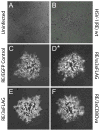
Comparison of the plaque phenotypes of isolated recombinant viruses to the parental HSV-1(RE) wildtype virus. Vero cell monolayers were mock infected (A) or infected with the parental HSV-1(RE) virus (B), the EGFP control virus that specified no immunological epitopes (C), the RE/ss3xFLAG virus (D), the RE/3xFLAG virus (E) or the RE/3xCD8Ova virus (F). 48 h p.i. viral plaques were visualized by phase and fluorescent microscopy and representative viral plaques were imaged and merged. (*) For the secretion signal containing 3x FLAG epitope virus, RE/ss3xFLAG (D) capture of the EGFP fluorescent image was twice as long as all other fluorescent exposures of viral plaques.
Figure 6
Figure 6
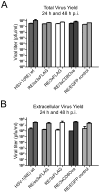
Comparison of total (A) and extracellular (B) viral yields by one step growth analysis. The parental HSV-1(RE) wildtype, RE/ss3xFLAG, RE/3xFLAG, RE/3xCD8 Ova and RE/EGFP control viruses were infected to Vero cells and at 24 and 48 h p.i. viral titers were determined from cell supernatants (extracellular) or cell lysates and supernatants (total).
Figure 7
Figure 7
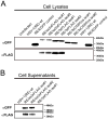
Detection of immunological epitope expression by recombinant viruses in infected cell lysates (A) and/or cell supernatants (B) by Western Blot analysis. The specific expression of immunological epitopes by the indicated recombinant viruses was indirectly detected (αGFP) by a shift in GFP’s apparent molecular mass (RE/EGFP control lane) caused by the fusion of the specified immunological epitope to the amino terminus of GFP. In addition, for the virus isolates that specified 3xFLAG immunological epitopes the presence of the FLAG epitopes were directly detected by an anti-FLAG monoclonal antibody (αFLAG).
Figure 8
Figure 8
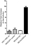
Quantitative assessment of OVA-specific CD8+ T cell responses to HSV-1(RE) viruses expressing immunological epitopes. MC57G fibroblasts were infected with either the wildtype HSV-1(RE) strain or the indicated immunological epitope expressing viruses. Infected cells were subsequently co-cultured with the LacZ-inducible, OVA/Kb-specific B3Z T cell hybridoma at an effector: target ratio of 1. Activation of B3Z T cells were quantified by a chemiluminescent beta-galactosidase assay and the fold induction of enzymatic activity was determined relative to wildtype HSV-1(RE) infected cells. The experiments were performed in triplicate and the mean +/− standard deviation is reported.

Similar articles

Cited by

References

    1. Barnden MJ, Allison J, Heath WR, Carbone FR. Defective TCR expression in transgenic mice constructed using cDNA-based alpha- and beta-chain genes under the control of heterologous regulatory elements. Immunol Cell Biol. 1998;76:34–40. - PubMed
    1. Bhattacharjee PS, Neumann DM, Foster TP, Clement C, Singh G, Thompson HW, Kaufman HE, Hill JM. Effective treatment of ocular HSK with a human apolipoprotein E mimetic peptide in a mouse eye model. Invest Ophthalmol Vis Sci. 2008;49:4263–8. - PubMed
    1. Brandt CR, Kolb AW, Shah DD, Pumfery AM, Kintner RL, Jaehnig E, Van Gompel JJ. Multiple determinants contribute to the virulence of HSV ocular and CNS infection and identification of serine 34 of the US1 gene as an ocular disease determinant. Invest Ophthalmol Vis Sci. 2003;44:2657–68. - PubMed
    1. Brandt CR. The role of viral and host genes in corneal infection with herpes simplex virus type 1. Exp Eye Res. 2005;80:607–21. - PubMed
    1. Cantin E, Chen J, Willey DE, Taylor JL, O’Brien WJ. Persistence of herpes simplex virus DNA in rabbit corneal cells. Invest Ophthalmol Vis Sci. 1992;33:2470–5. - PubMed

Publication types

MeSH terms

Substances

LinkOut - more resources