. 2012 Jan 1;361(1):103-15.
doi: 10.1016/j.ydbio.2011.10.017. Epub 2011 Oct 18.

Negative regulation of Yap during neuronal differentiation

Affiliations

Negative regulation of Yap during neuronal differentiation

Huanqing Zhang et al. Dev Biol. .

Abstract

Regulated proliferation and cell cycle exit are essential aspects of neurogenesis. The Yap transcriptional coactivator controls proliferation in a variety of tissues during development, and this activity is negatively regulated by kinases in the Hippo signaling pathway. We find that Yap is expressed in mitotic mouse retinal progenitors and it is downregulated during neuronal differentiation. Forced expression of Yap prolongs proliferation in the postnatal mouse retina, whereas inhibition of Yap by RNA interference (RNAi) decreases proliferation and increases differentiation. We show Yap is subject to post-translational inhibition in the retina, and also downregulated at the level of mRNA expression. Using a cell culture model, we find that expression of the proneural basic helix-loop-helix (bHLH) transcription factors Neurog2 or Ascl1 downregulates Yap mRNA levels, and simultaneously inhibits Yap protein via activation of the Lats1 and/or Lats2 kinases. Conversely, overexpression of Yap prevents proneural bHLH proteins from initiating cell cycle exit. We propose that mutual inhibition between proneural bHLH proteins and Yap is an important regulator of proliferation and cell cycle exit during mammalian neurogenesis.


Figures

Figure 1
Figure 1
Yap expression and Hippo pathway activation in neonatal mouse retina. In situ hybridization (A–C) or indirect immunofluorescence (D–F) on P0 retina sections. (A, B) Elavl3 (HuC) mRNA, a marker for differentiated neurons (Okano and Darnell, 1997), is detected in the inner nuclear layer (INL) and ganglion cell layer (GCL), while BrdU labels S-phase cells in the neuroblast layer (NBL). (C) Yap mRNA is expressed primarily in the NBL. (D) Yap protein is present in nuclei in the NBL, and overlaps with S-phase cells labeled by BrdU (E, F). (G) Yap mRNA, measured by qRT-PCR, decreases between P0 and P7 in the developing retina. (H) By western blot, Yap protein decreases at P2 and later, as retinal neurogenesis decreases in the postnatal retina, while phospho-Mst1/2 (p-Mst1/2) and phospo-Lats1/2 (p-Lats1/2) increase. Yap is phosphorylated at S112 (p-Yap) in the retina. β-actin protein was used as a loading control.
Figure 2
Figure 2
Yap RNAi reduces cell proliferation and increases differentiation in postnatal mouse retinas. (A) Schematic diagrams of UI4-GFP-SIBR vectors expressing 2 tandem copies of the same shRNA against luciferase (luc) or Yap mRNA. Numbers in shRNA names indicate the starting base of the target sequence in the mRNA. (B,D) Mouse retinas were electroporated in vivo with two different shRNA vectors against Yap mRNA or the luc control shRNA vector at P0. (B) At P2.5, retinas were collected after labeling with BrdU for 2 hours. Sections were stained with an anti-BrdU antibody and transfected cells were identified by GFP fluorescence. (C) Quantitation of BrdU positive cells among GFP positive cells, based on 6 sections from each retina (mean ± S.D., n = 5 retinas). (D) At P2.5, retina sections were stained with an anti-β-tubulin III antibody and transfected cells were identified by GFP fluorescence. (E) Quantitation of β-tubulin III positive cells among GFP positive cells, based on 3 sections from each retina (mean ± S.D., n = 3 retinas). Scale bar: 50µm **p<0.01. IPL: inner plexiform layer; other labels as in Fig.1.
Figure 3
Figure 3
Yap promotes cell proliferation in mouse retinas. (A) Schematic diagrams of plasm id DNA vectors coexpressing GFP and wild-type mouse Yap, the phosphorylation site mutant YapS112A, the activation domain deletion YapΔC, or a multimerized Myc epitope tag (MT) as a control. (B) Plasmid DNA vectors introduced into retinas by in vivo electroporation at P0. At P7, BrdU was administered 2 hours before harvesting retinas. Sections were stained with an anti-BrdU antibody and transfected cells were identified using GFP. In the MT control, nearly all transfected retinal cells were BrdU negative. In contrast, a subset of Yap vector transfected cells were BrdU positive, and the retinal layers were slightly disorganized including clusters of GFP-positive cell bodies in the IPL. Retinal layering was disrupted and more BrdU/GFP double labeled cells were present with the Yap S112A mutant. In the YapΔC retina, almost all transfected cells were BrdU negative. Scale bar: 50µm. (C) Quantitation of BrdU positive cells among GFP positive cells based on 6 sections from each retina. Data is mean ± S.D. (n = 4 retinas). In this and subsequent figures: *p<0.05, **p<0.01. ONL: outer nuclear layer. Other labels as in Fig.2.
Figure 4
Figure 4
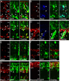
Characterization of retinal cells overexpressing Yap. (A–H) Mouse retinas electroporated in vivo with the Yap/GFP vector at P0 and fixed at P7 were labeled with various antibodies (see text). (A) Arrowheads indicate GFP-positive cells expressing the proliferation marker Ki-67. Arrow: GFP-negative cell labeled by Ki-67. (B) Arrowheads indicate GFP-positive cells expressing Sox2 that incorporated BrdU. (C) Arrowheads indicate cells expressing GFP and Sox9, a marker of retinal progenitors and Müller glia. (D) Arrowheads denote cells expressing GFP, Pax6, and Sox2, proteins expressed in retinal progenitor cells and amacrine neurons. (E) Islet1, a marker of amacrine and ganglion cells, does not overlap with GFP (F) AP2α, a pan-amacrine marker, does not overlap with GFP. (G) Arrowheads denote presumptive bipolar cells expressing GFP and Prox1. (H) Arrowheads indicate cells expressing GFP and the photoreceptor protein Recoverin.
Figure 5
Figure 5
Neurog2 or Ascl1 downregulate Yap mRNA and protein in P19 cells; Yap regulates cell cycle exit during neuronal differentiation of P19 cells driven by Neurog2. (A,B) P19 cells were transiently transfected with MT control, Neurog2, or Ascl1 expression vectors, and a puroR (puromycin resistance) vector. Transfected cells were selected with puromycin prior to RNA collection or lysis. (A) Yap mRNA levels were determined 24 and 48 hrs after transfection by qRT-PCR. Normalized data are mean ± S.D. (n = 3). (B) Endogenous Yap and p-Yap levels in transfected cells were analyzed by western blot. ERK protein was used as a loading control. (C) Overexpression of Yap drives P19 cells expressing Neurog2 to continue proliferation. Plasmid DNA vectors coexpressing GFP and wild-type mouse Yap, YapΔC, or the MT control were co-transfected with Neurog2 into P19 cells. Cells were pulse-labeled with BrdU at indicated times followed by staining with anti-BrdU antibody. Transfected cells were identified by GFP expression. The percentage of BrdU positive cells among GFP positive cells was quantified from three independent transfections at each condition. (D) Yap RNAi increases cell cycle exit in P19 cells transfected with Neurog2. Cells co-transfected with Neurog2 and RNAi vectors against Yap or a control RNAi vector (luc) were pulse-labeled with BrdU followed by staining with anti-BrdU antibody. The BrdU-positive fraction of GFP transfected cells was determined from three independent transfections at each condition (mean ± S.D. for n = 3). (E) Yap overexpression does not prevent upregulation of the endogenous Hes6 mRNA by Neurog2. Plasmid vectors for Neurog2 or control protein MT were transfected with or without Yap in P19 cells. Hes6 mRNA levels determined by qRT-PCR 48 hours after transfection.
Figure 6
Figure 6
Yap nuclear localization is regulated by expression of Neurog2 or Ascl1. P19 cells were cotransfected with the MT-Yap or MT-YapS112A expression vectors, Neurog2, Ascl1, or an empty vector control as indicated, and the puroR vector. MT-Yap or MT-YapS112A were detected by indirect immunofluorescence with an anti-Myc antibody; nuclei were stained with DAPI at 48 hr after transfection. (A) MT-Yap is detected in both cytoplasm and nuclei in the control cells, but expression of Neurog2 or Ascl1 leads to nuclear exclusion of MT-Yap. (B) Neurog2 or Ascl1 expression does not lead to nuclear exclusion of MT-YapS112A. Arrowheads indicate representative nuclei. Scale bar:10µm.
Figure 7
Figure 7
Neurog2 and Ascl1 activate the Lats1/2 kinases; Lats1/2 RNAi regulates endogenous Yap protein and decreases proliferation during neuronal differentiation of P19 cells. (A) Western blot for p-Lats1/2 and p-Mst1/2 in P19 cells transfected with Neurog2, Ascl1 or control MT expression vectors. Actin was used as a loading control. (B) Schematic diagrams of UI4-GFP-SIBR vectors expressing 4 tandem copies of the same shRNA against luciferase mRNA (control), or 2 tandem copies each of shRNAs targeting Lats1 and Lats2 mRNA. (C) Lats1/2 RNAi decreases nuclear exclusion of MT-Yap in P19 cells expressing Neurog2. P19 cells were cotransfected with MT-Yap or MT-YapS112A expression vectors and a Neurog2 expression vector or control empty vector. Cells were stained with anti-Myc antibody and nuclei were stained with DAPI 48 hr after transfection. Arrowheads indicate representative nuclei. Scale bar:10µm. (D, E) P19 cells were transiently cotransfected with Lats1/2 shRNA vectors or a control shRNA vector, the Neurog2 expression vector, and the puroR vectors; transfected cells were selected with puromycin. (D) RNAi against Lats1and Lats2 increases endogenous nuclear Yap. Nuclear and cytoplasmic protein fractions were prepared and analyzed by western blot with anti-Yap antibody. Actin and Histone H3 were used as loading controls for cytoplasmic and nuclear proteins respectively. (E) Endogenous total Yap and p-Yap were determined by western blot at 48 hr after transfection. Actin was used as a loading control. Bar graph shows the fold change of total Yap or phosphorylated Yap quantified by densitometry. Data is mean ± S.D (n=3). (F) Lats1/2 RNAi leads to increased cell proliferation in P19 cells expressing Neurog2. P19 cells were cotransfected with a Neurog2 expression vector, Lats1/2 or control shRNA vectors (GFP), and either a Lats2 expression vector (white bar) or the control MT vector (grey bar). Cells were pulse labeled with BrdU followed by immunostaining with anti-BrdU antibody. The percentage of BrdU positive cells among GFP positive cells was quantified from three independent transfections at each condition. Data is mean ± S.D.
Figure 8
Figure 8
Schematic diagram of mutual inhibition between Yap and Neurog2. Yap can inhibit cell cycle exit and neuronal differentiation in Neurog2 expressing cells, while Neurog2 indirectly inhibits Yap function via activation of the Lats1/2 kinases and by downregulation of Yap mRNA expression. Neurog2 may activate Lats1/2 via the Mst1/2 kinases (see Discussion). Yap also may inhibit apoptosis, as has been observed in other systems.

References

    1. Akagi T, Inoue T, Miyoshi G, Bessho Y, Takahashi M, Lee JE, Guillemot F, Kageyama R. Requirement of multiple basic helix-loop-helix genes for retinal neuronal subtype specification. J Biol Chem. 2004;279:28492–28498. - PubMed
    1. Asthagiri AR, Parry DM, Butman JA, Kim HJ, Tsilou ET, Zhuang Z, Lonser RR. Neurofibromatosis type 2. Lancet. 2009;373:1974–1986. - PMC - PubMed
    1. Bassett EA, Pontoriero GF, Feng W, Marquardt T, Fini ME, Williams T, West-Mays JA. Conditional deletion of activating protein 2alpha (AP-2alpha) in the developing retina demonstrates non-cell-autonomous roles for AP-2alpha in optic cup development. Mol Cell Biol. 2007;27:7497–7510. - PMC - PubMed
    1. Basu S, Totty NF, Irwin MS, Sudol M, Downward J. Akt phosphorylates the Yes-associated protein, YAP, to induce interaction with 14-3-3 and attenuation of p73-mediated apoptosis. Mol Cell. 2003;11:11–23. - PubMed
    1. Ben-Arie N, Bellen HJ, Armstrong DL, McCall AE, Gordadze PR, Guo Q, Matzuk MM, Zoghbi HY. Math1 is essential for genesis of cerebellar granule neurons. Nature. 1997;390:169–172. - PubMed

Publication types

MeSH terms