. 2012 Mar;25(3):416-33.
doi: 10.1038/modpathol.2011.166. Epub 2011 Oct 28.

The distribution of immunomodulatory cells in the lungs of patients with idiopathic pulmonary fibrosis

Affiliations

The distribution of immunomodulatory cells in the lungs of patients with idiopathic pulmonary fibrosis

Gerard J Nuovo et al. Mod Pathol. 2012 Mar.

Abstract

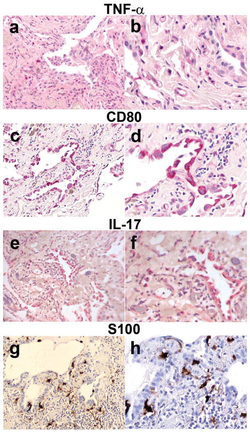
We have characterized the immune system involvement in the disease processes of idiopathic pulmonary fibrosis in novel ways. To do so, we analyzed lung tissue from 21 cases of idiopathic pulmonary fibrosis and 21 (non-fibrotic, non-cancerous) controls for immune cell and inflammation-related markers. The immunohistochemical analysis of the tissue was grouped by patterns of severity in disease pathology. There were significantly greater numbers of CD68(+) and CD80(+) cells and significantly fewer CD3(+), CD4(+), and CD45RO(+) cells in areas of relatively (histologically) normal lung in biopsy samples from idiopathic pulmonary fibrosis patients compared with controls. In zones of active disease, characterized by epithelial cell regeneration and fibrosis, there were significantly more cells expressing CD4, CD8, CD20, CD68, CD80, chemokine receptor 6 (CCR6), S100, IL-17, tumor necrosis factor-α, and retinoic acid-related orphan receptors compared with histologically normal lung areas from idiopathic pulmonary fibrosis patients. Inflammation was implicated in these active regions by the cells that expressed retinoid orphan receptor-α, -β, and -γ, CCR6, and IL-17. The regenerating epithelial cells predominantly expressed these pro-inflammatory molecules, as evidenced by co-expression analyses with epithelial cytokeratins. Macrophages in pseudo-alveoli and CD3(+) T cells in the fibrotic interstitium also expressed IL-17. Co-expression of IL-17 with retinoid orphan receptors and epithelial cytoskeletal proteins, CD68, and CD3 in epithelial cells, macrophages, and T-cells, respectively, confirmed the production of IL-17 by these cell types. There was little staining for forkhead box p3, CD56, or CD34 in any idiopathic pulmonary fibrosis lung regions. The fibrotic regions had fewer immune cells overall. In summary, our study shows participation of innate and adaptive mononuclear cells in active-disease regions of idiopathic pulmonary fibrosis lung, where the regenerating epithelial cells appear to propagate inflammation. The regenerative mechanisms become skewed to ultimately result in lethal, fibrotic restriction of lung function.


Conflict of interest statement

Conflict of Interest

The authors have no conflicts of interest to disclose.

Figures

Figure 1
Figure 1
Lymphocytes, Monocytes, and Macrophages The mean ± standard error of the mean (SEM) of the numbers of cells stained positive for the labeled surface antigens on slides with lung tissue from the indicated idiopathic pulmonary fibrosis signature histological regions and control lung tissue. Asterisks indicate the p-values for comparison of the idiopathic pulmonary fibrosis tissues to the control lung tissue (*p < 0.05; **p < 0.005; ***p < 0.0005). Deltas indicate the p values for comparison of the differences involving the data bars that they appear between (δp < 0.05; δδp < 0.005; δδδp < 0.0005).
Figure 2
Figure 2
Cell Markers and Cytokines The mean ±SEM of the numbers of cells stained positive for the indicated antigens, chemokine receptors, or cytokines on slides with lung tissue from the indicated idiopathic pulmonary fibrosis signature histological regions or control lung tissue. Asterisks indicate the p-values for comparison of the idiopathic pulmonary fibrosis tissues to the control lung tissue (*p < 0.05; **p < 0.005; ***p < 0.0005). Deltas indicate the p values for comparison of the differences involving the data bars that they appear between (δp < 0.05; δδp < 0.005; δδδp < 0.0005).
Figure 3
Figure 3
Control Lung, Idiopathic Pulmonary Fibrosis-Normal Lung, and Idiopathic Pulmonary Fibrosis-Epithelial Dominant Lung 3a, 3d, 3g, 3j, and 3m show lung tissue from the controls probed for the indicated cellular markers; 3b, 3e, 3h, 3k, and 3n show the staining of histologically normal lung from idiopathic pulmonary fibrosis patients; and 3c, 3f, 3i, 3l, and 3o show the staining of the epithelial dominant or active phase of idiopathic pulmonary fibrosis. All photomicrographs show the tissue with a magnification of 200X. The tissues shown in photos 3a–3f were stained with diaminobenzidine; the tissues in photos 3g–3o were stained with Fast Red. The counterstain in each case was hematoxylin.
Figure 4
Figure 4
Idiopathic Pulmonary Fibrosis Inflammatory Infiltrates All photomicrographs show the tissue stained with Fast Red and hematoxylin counterstain. The photos in 4a–4f have a magnification of 200X; 4g and 4h are at 400X.
Figure 5
Figure 5
Idiopathic Pulmonary Fibrosis-Epithelial Dominant Areas 5a–5f show idiopathic pulmonary fibrosis lung tissue stained with Fast Red and hematoxylin counterstain, and 5g and 5h show idiopathic pulmonary fibrosis lung tissue stained with diaminobenzidine and hematoxylin counterstain. 5a, 5c, 5e, and 5g have a magnification of 100X, the others have a magnification of 400X.
Figure 6
Figure 6
Co-expression Analyses of Epithelial Cytokeratins, CCR6 and IL-17 in the Idiopathic Pulmonary Fibrosis-Epithelial Dominant Areas. The images in 6a–6d are photos of the same slide, as are 6e and f. 6a shows the color-based image where the bound cytokeratin AE1/3 Ab appears brown (diaminobenzidine), CCR6 is red (Fast Red), and the counterstain (visible on nuclei) is blue (hematoxylin). The Nuance system converts diaminobenzidine to fluorescent green showing the cytokeratins in 6b. The CCR6 and IL-17 (Fast Red) are converted to fluorescent red (6c and 6e), fluorescent yellow marks cells with both targets (6d and 6f). The CCR6+ cells co-express cytokeratins (6d). Co-expression analyses with IL 17 (6f) shows that the epithelial IL 17+ cells are cytokeratin AE1/3 Ab+, but many IL-17+/cytokeratin AE1/3 cells are fluorescent red in the stroma.
Figure 7
Figure 7
Co-expression Analyses of IL-17 with CD3 and CD68 in the Idiopathic Pulmonary Fibrosis-Epithelial Dominant Stage. 7a shows the color-based image where the CD3 is stained brown (diaminobenzidine), IL-17 is red (Fast Red), and the counterstain is blue (hematoxylin). 7b shows the same image made fluorescent by the Nuance system. Some double-positive (yellow) stained cells are present in the interstitium (Th-17s), and some of the CD3+ T cells are green, both have the morphology of lymphocytes. Fast Red-labeled cells (IL-17+, non-T cells) are also apparent. 7c shows the regular color-based image with CD68+ stained brown (diaminobenzidine) and the IL-17 is again stained with Fast Red. 7d shows the same image made fluorescent, with double-positive macrophages (yellow), some green macrophages (CD68+) in the interstitium, and red fluorescent (IL-17+) regenerating epithelial cells.
Figure 8
Figure 8
Intracellular Retinoid-like Orphan Receptor-Positive Cells The mean ± SEM of the numbers of interstitial cells stained positive for the ROR antigens on slides with areas of lung tissue representing the signature idiopathic pulmonary fibrosis histological regions and control lung tissue. Asterisks indicate the p-values for comparison of the idiopathic pulmonary fibrosis tissues to the control lung tissue (*p < 0.05; **p < 0.005; ***p < 0.0005). Deltas indicate the p values for comparison of the differences involving the data bars that they appear between (δp < 0.05; δδp < 0.005; δδδp < 0.0005).
Figure 9
Figure 9
Co-expression Analyses of RORs and Cytokeratins in the Idiopathic Pulmonary Fibrosis-Epithelial Dominant Areas. 9a, 9b and 9c show the regular color-based images where the cytokeratin AE1/3 Ab is brown (diaminobenzidine); ROR-α, -β, and -γ are Fast Red labeled; and the counterstain is blue (hematoxylin). The same images (arranged vertically) are fluorescence-converted by the Nuance system in photos 9d–9l. Most of the ROR-expressing cells are stained positive with the cytokeratin AE1/3 Ab. Control lung tissues were double stained with the same antibodies and color reagents, the co-expression is shown and labeled in 9m, 9n, and 9o. Green fluorescence of the cytokeratin AE1/3 Ab can be seen, but co-expression with the RORs is absent.

References

    1. Olsen AL, Swigris JJ, Lezotte DC, Norris JM, Wilson CG, Brown KK. Mortality from pulmonary fibrosis increased in the United States from 1992 to 2003. Am J Respir Crit Care Med. 2007;176:277–84. - PubMed
    1. Raghu G, Collard HR, Egan JJ, Martinez FJ, Behr J, Brown KK, et al. American Thoracic Society Documents. An official ATS/ERS/JRS/ALAT statement: Idiopathic pulmonary fibrosis: Evidence-based guidelines for diagnosis and management. Am J Respir Crit Care Med. 2011;183:788–824. - PMC - PubMed
    1. Katzenstein A-LA, Mukhopadhyay S, Myers JL. Diagnosis of usual interstitial pneumonia and distinction from other fibrosing interstitial lung diseases. Hum Pathol. 2008;39:1562–81. - PubMed
    1. Chapman HA. Disorders of lung matrix remodeling. J Clin Invest. 2004;113:148–57. - PMC - PubMed
    1. Selman M, Pardo A, Kaminski N. Idiopathic pulmonary fibrosis: Aberrant recapitulation of developmental programs? PLoS Medicine. 2008;5:e62. - PMC - PubMed

Publication types

MeSH terms