

Atomic-resolution dynamics on the surface of amyloid-β protofibrils probed by solution NMR
- PMID: 22037310
- PMCID: PMC3237923
- DOI: 10.1038/nature10577
Atomic-resolution dynamics on the surface of amyloid-β protofibrils probed by solution NMR
Abstract
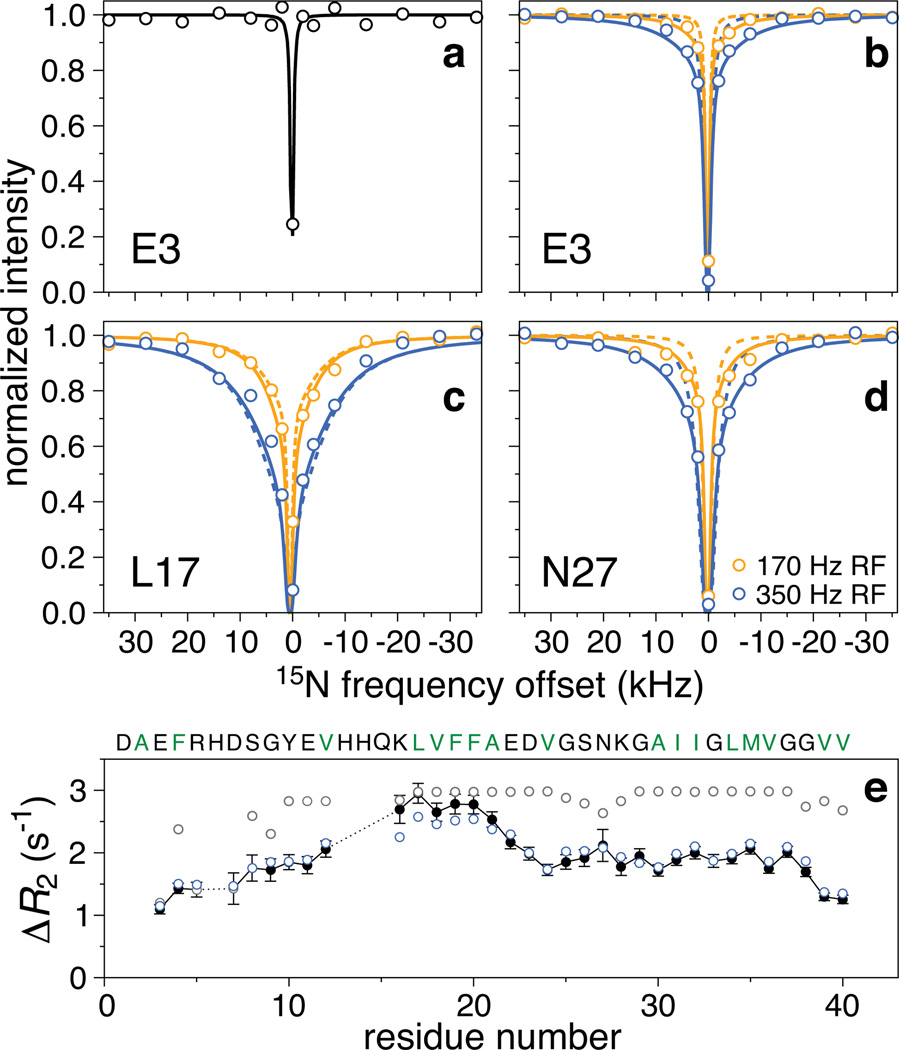
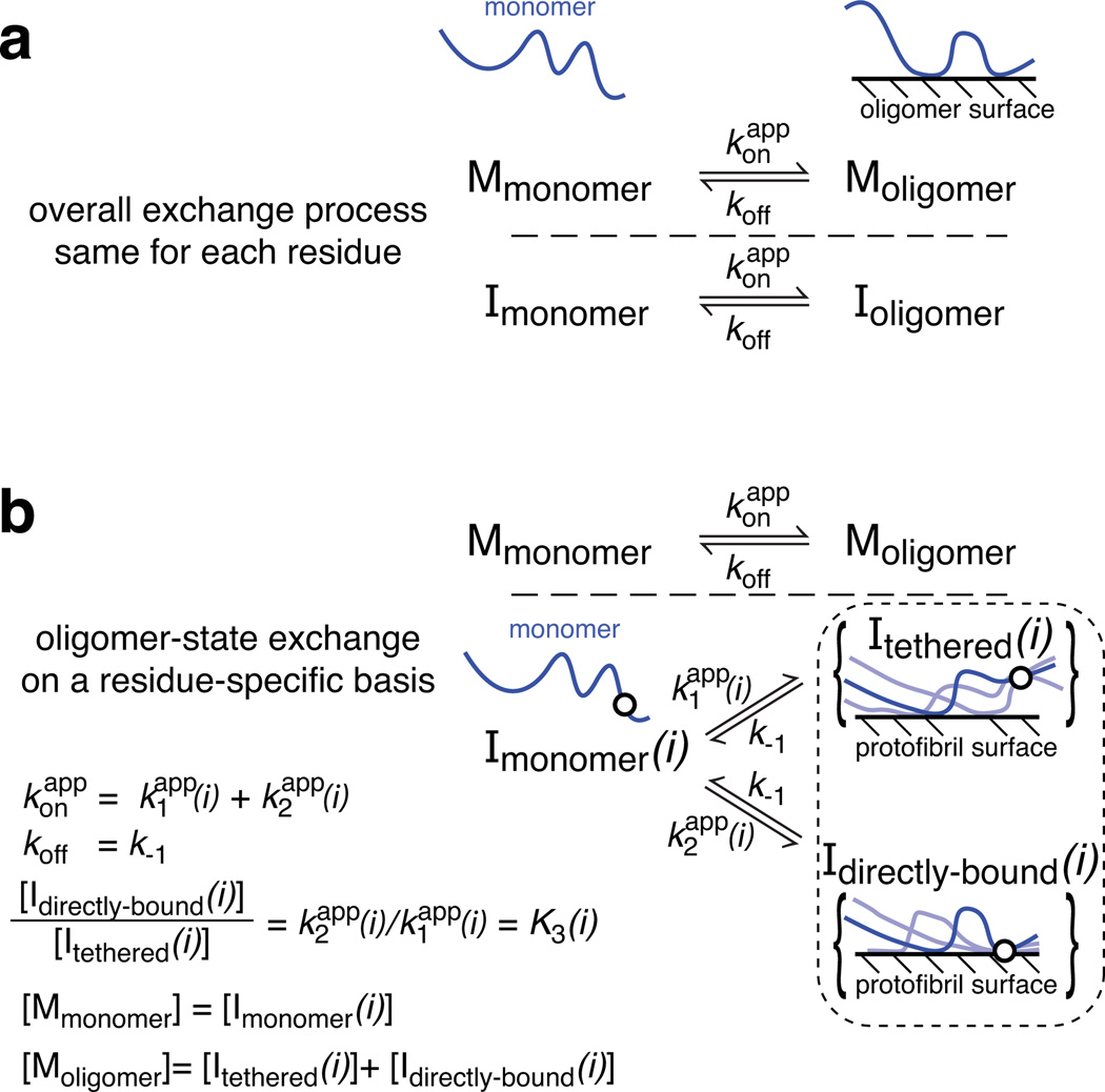
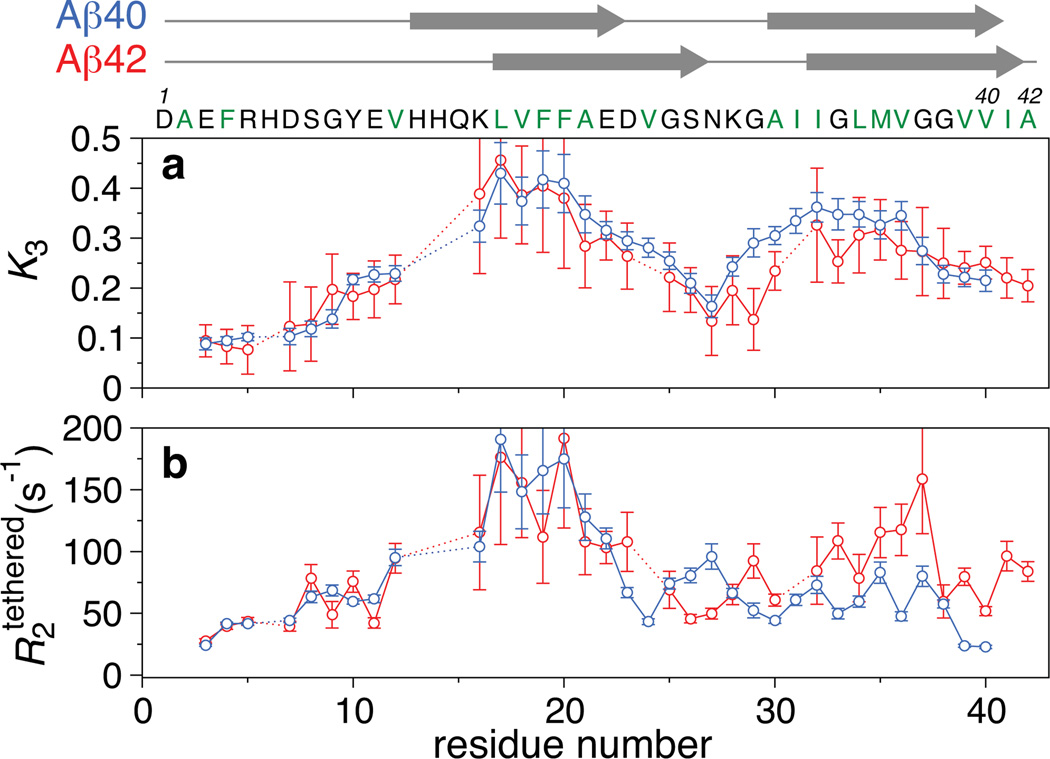
Exchange dynamics between molecules free in solution and bound to the surface of a large supramolecular structure, a polymer, a membrane or solid support are important in many phenomena in biology and materials science. Here we present a novel and generally applicable solution NMR technique, known as dark-state exchange saturation transfer (DEST), to probe such exchange phenomena with atomic resolution. This is illustrated by the exchange reaction between amyloid-β (Aβ) monomers and polydisperse, NMR-invisible ('dark') protofibrils, a process of significant interest because the accumulation of toxic, aggregated forms of Aβ, from small oligomers to very large assemblies, has been implicated in the aetiology of Alzheimer's disease. The (15)N-DEST experiment imprints with single-residue-resolution dynamic information on the protofibril-bound species in the form of (15)N transverse relaxation rates ((15)N-R(2)) and exchange kinetics between monomers and protofibrils onto the easily observed two-dimensional (1)H-(15)N correlation spectrum of the monomer. The exchanging species on the protofibril surface comprise an ensemble of sparsely populated states where each residue is either tethered to (through other residues) or in direct contact with the surface. The first eight residues exist predominantly in a mobile tethered state, whereas the largely hydrophobic central region and part of the carboxy (C)-terminal hydrophobic region are in direct contact with the protofibril surface for a significant proportion of the time. The C-terminal residues of both Aβ40 and Aβ42 display lower affinity for the protofibril surface, indicating that they are likely to be surface exposed rather than buried as in structures of Aβ fibrils, and might therefore comprise the critical nucleus for fibril formation. The values, however, are significantly larger for the C-terminal residues of Aβ42 than Aβ40, which might explain the former's higher propensity for rapid aggregation and fibril formation.
Figures
References
-
- Lashuel HA, Lansbury PT. Are amyloid diseases caused by protein aggregates that mimic bacterial pore-forming toxins? Q. Rev. Biophys. 2006;39:167–201. - PubMed
-
- Walsh DM, Selkoe DJ. A beta Oligomers - a decade of discovery. J. Neurochem. 2007;101:1172–1184. - PubMed
-
- Querfurth HW, LaFerla FM. Mechanisms of Disease Alzheimer's Disease. New Eng. J. Med. 2010;362:329–344. - PubMed
Publication types
MeSH terms
Substances
Grants and funding
LinkOut - more resources
Full Text Sources
