. 2012 Jan;233(1):243-52.
doi: 10.1016/j.expneurol.2011.10.009. Epub 2011 Oct 19.

Neuronal activity and axonal sprouting differentially regulate CNTF and CNTF receptor complex in the rat supraoptic nucleus

Affiliations

Neuronal activity and axonal sprouting differentially regulate CNTF and CNTF receptor complex in the rat supraoptic nucleus

Jason M Askvig et al. Exp Neurol. 2012 Jan.

Abstract

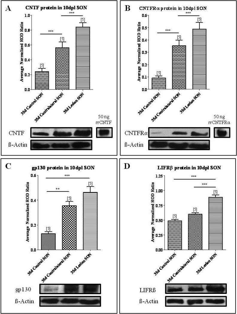
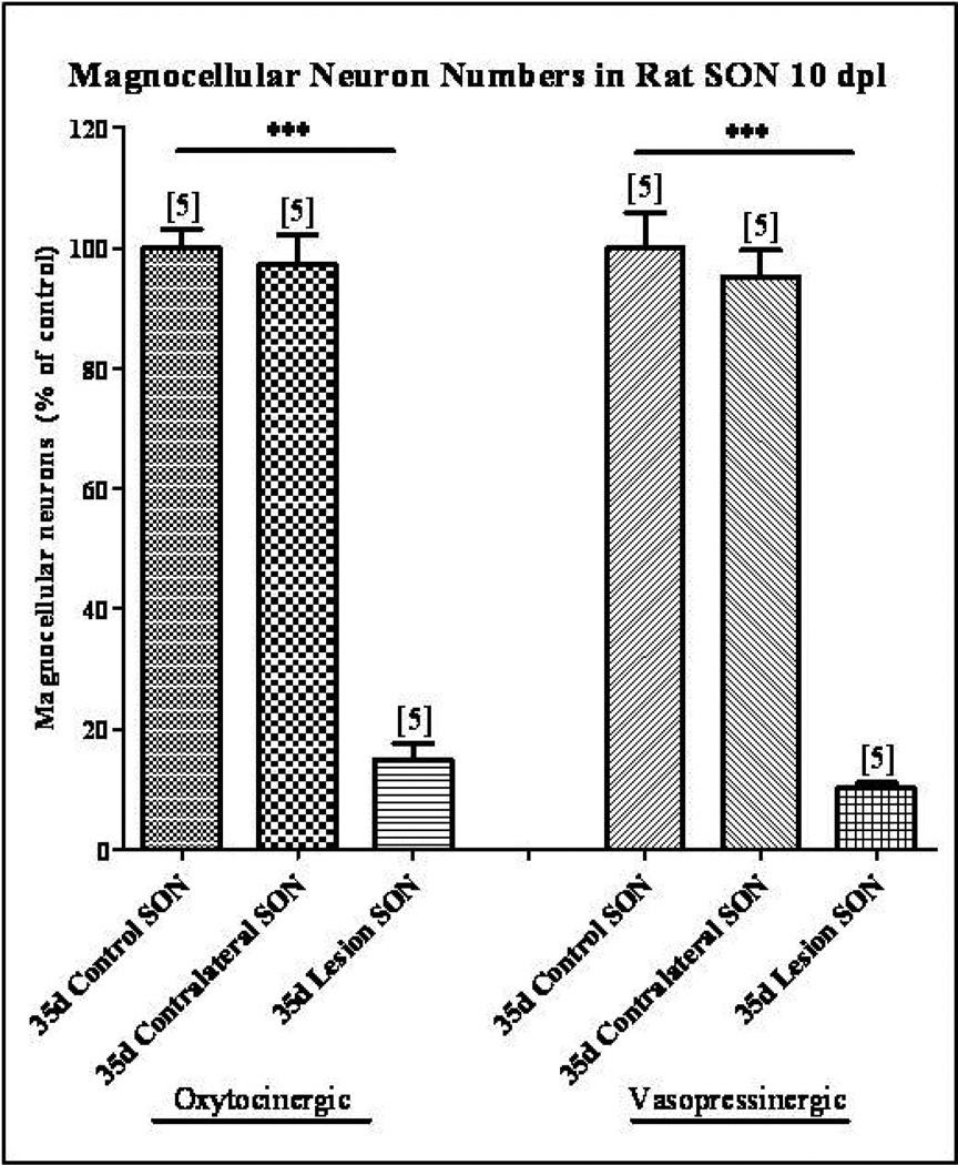
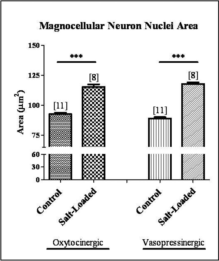
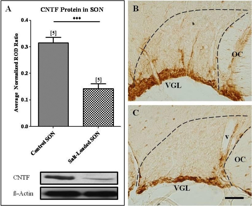
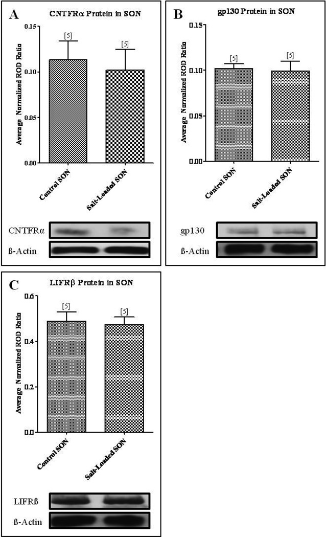
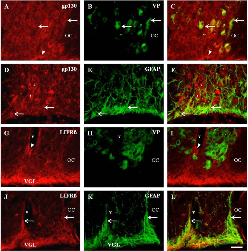
We demonstrated previously that the hypothalamic supraoptic nucleus (SON) undergoes a robust axonal sprouting response following unilateral transection of the hypothalamo-neurohypophysial tract. Concomitant with this response is an increase in ciliary neurotrophic factor (CNTF) and CNTF receptor alpha (CNTFRα) expression in the contralateral non-uninjured SON from which the axonal outgrowth occurs. While these findings suggest that CNTF may act as a growth factor in support of neuronal plasticity in the SON, it remained to be determined if the observed increase in neurotrophin expression was related to the sprouting response per se or more generally to the increased neurosecretory activity associated with the post-lesion response. Therefore we used immunocytochemistry and Western blot analysis to examine the expression of CNTF and the components of the CNTF receptor complex in sprouting versus osmotically-stimulated SON. Western blot analysis revealed a significant increase in CNTF, CNTFRα, and gp130, but not LIFRß, protein levels in the sprouting SON at 10days post lesion in the absence of neuronal loss. In contrast, osmotic stimulation of neurosecretory activity in the absence of injury resulted in a significant decrease in CNTF protein levels with no change in CNTFRα, gp130, or LIFRß protein levels. Immunocytochemical analysis further demonstrated gp130 localization on magnocellular neurons and astrocytes while the LIFRß receptor was found only on astrocytes in the SON. These results are consistent with the hypothesis that increased CNTF and CNTFR complex in the sprouting, metabolically active SON are related directly to the sprouting response and not the increase in neurosecretory activity.


Figures

Fig. 1
Fig. 1. CNTF and CNTF receptor complex are increased following axotomy
Western blot analysis demonstrated a significant increase in CNTF (A), CNTFRα (B), gp130 (C), and LIFRβ (D) protein levels in the axotomized SON compared to age-matched control. Within the sprouting SON contralateral to the lesion, CNTF (A), CNTFRα (B), and gp130 (C) protein levels were significantly increased compared to age-matched control. However, LIFRβ levels were not significantly changed. Column bars and error bars represent the mean and SEM of 5 groups. Each group represents isolated SON pooled from six rats. **p<0.01, ***p<0.0001.
Fig. 2
Fig. 2. Magnocellular neuron survival following unilateral lesion
Immunohistochemical labeling of OT and VP neurons was used to identify individual magnocellular neurons. Cell counts demonstrated no significant decrease in the number of OT or VP neurons in the SON contralateral to the unilateral lesion. However, at 10 dpl the numbers of OT and VP neurons were reduced by 85% and 90% respectively in the axotomized SON. Column bars and error bars represent the mean and SEM of each group. Each group is comprised of a minimum of six sections sampled from each of 5 animals. ***p<0.0001.
Fig. 3
Fig. 3. Chronic salt-loading activates magnocellular neurons in the SON
Immunocytochemical analysis demonstrated a significant hypertrophy of 25% and 32% in OT and VP magnocellular neuron nuclei area, respectively, in the chronic salt-loaded SON. Column bars and error bars represent the mean and SEM of each group. ***p<0.0001.
Fig. 4
Fig. 4. Physiological activation of the SON decreased CNTF protein levels in the SON
(A) Western blot analysis revealed a significant decrease in CNTF protein levels in the salt-loaded SON compared to the age-matched control SON. Immunocytochemical analysis confirmed these results. A decrease in CNTF-immunoreactivity in the SON of the physiologically activated SON (C) was apparent when compared to the age-matched control SON (B). Column bars and error bars represent the mean and SEM of each group. OC, optic chiasm; v, blood vessel; VGL, ventral glial limitans. Scale bar = 100 µm. ***p<0.0001.
Fig. 5
Fig. 5. Physiological activation of the SON resulted in no change in protein levels of the CNTF receptor components
Western blot analysis revealed no change in CNTFRα (A), gp130 (B), and LIFRβ (C) protein levels in the salt-loaded SON compared to the age-matched control SON. Column bars and error bars represent the mean and SEM of each group.
Fig. 6
Fig. 6. Differential expression of CNTF receptor complex on astrocytes and magnocellular neurons
Dual fluorescent colocalization of anti-gp130 (A), with anti-VP (B), revealed colocalization in vasopressinergic neurons (C, arrows). Note the gp130-immunoreactive profiles in presumptive astrocytes in the ventral glial limitans (VGL) of the SON (A, C, arrowheads). Similar observations were observed with anti-OT (not shown). Immunocytochemical analysis also demonstrated colocalization of anti-gp130 (D), with GFAP-immunoreactive astrocytes (E), of the SON (F, arrows). Also present are presumptive magnocellular neurons that are immunopositive for gp130 (D, F, asterisks). Unlike gp130, there was no observable colocalization of anti-LIFRβ in the magnocellular neurons of the SON (G–I). Note the LIFRβ-immunoreactive profiles surrounding blood vessels (v) in the SON (G, I, arrowheads) which do not colocalize with anti-VP (H) or anti-OT (not shown). However, strong LIFRβ-immunoreactivity in the ventral glial limitans (VGL) of the SON (J), revealed extensive colocalization of anti-LIFRβ (J), with anti-GFAP (K), throughout the entire SON (L, arrows). OC, optic chiasm; OT, oxytocin; v, blood vessel; VGL, ventral glial limitans; VP, vasopressin. Scale bar = 50 µm.

References

    1. Albrecht PJ, Dahl JP, Stoltzfus OK, Levenson R, Levison SW. Ciliary neurotrophic factor activates spinal cord astrocytes, stimulating their production and release of fibroblast growth factor-2, to increase motor neuron survival. Exp Neurol. 2002;173:46–62. - PubMed
    1. Aliaga E, Arancibia S, Givalois L, Tapia-Arancibia L. Osmotic stress increases brain-derived neurotrophic factor messenger RNA expression in the hypothalamic supraoptic nucleus with differential regulation of its transcripts. Relation to arginine-vasopressin content. Neuroscience. 2002;112:841–850. - PubMed
    1. Arakawa Y, Sendtner M, Thoenen H. Survival effect of ciliary neurotrophic factor (CNTF) on chick embryonic motoneurons in culture: comparison with other neurotrophic factors and cytokines. J Neurosci. 1990;10:3507–3515. - PMC - PubMed
    1. Arancibia S, Lecomte A, Silhol M, Aliaga E, Tapia-Arancibia L. In vivo brain-derived neurotrophic factor release and tyrosine kinase B receptor expression in the supraoptic nucleus after osmotic stress stimulus in rats. Neuroscience. 2007;146:864–873. - PubMed
    1. Armstrong WE, Gallagher MJ, Sladek CD. Noradrenergic stimulation of supraoptic neuronal activity and vasopressin release in vitro: mediation by an alpha 1-receptor. Brain Res. 1986;365:192–197. - PubMed

Publication types

MeSH terms