. 2011 Oct 30;14(12):1517-24.
doi: 10.1038/nn.2950.

Modulation of dADAR-dependent RNA editing by the Drosophila fragile X mental retardation protein

Affiliations

Modulation of dADAR-dependent RNA editing by the Drosophila fragile X mental retardation protein

Balpreet Bhogal et al. Nat Neurosci. .

Abstract

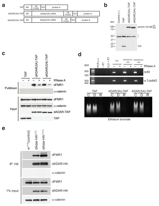
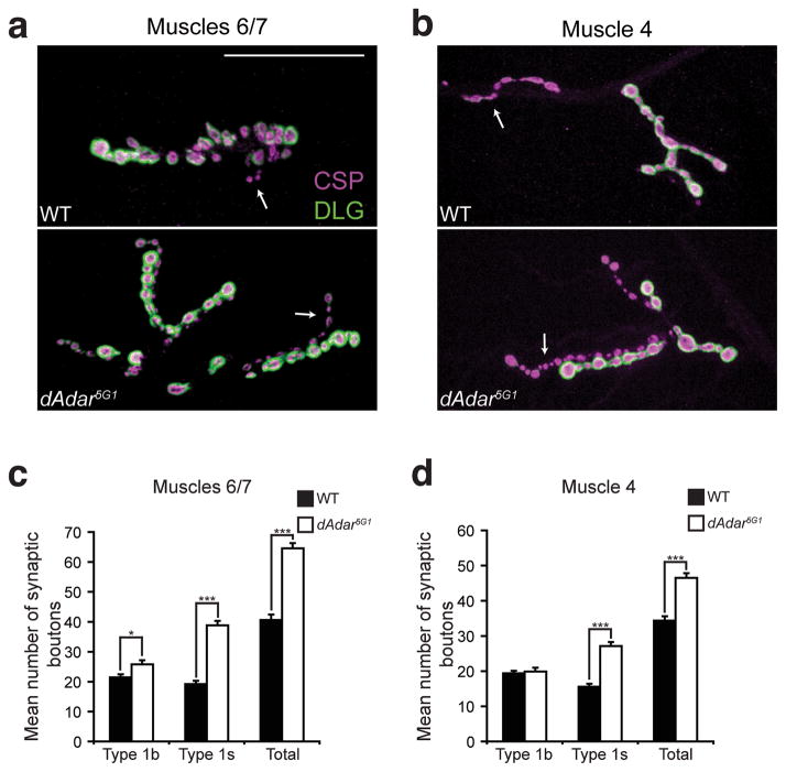
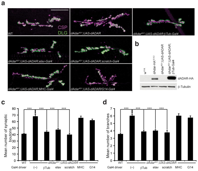
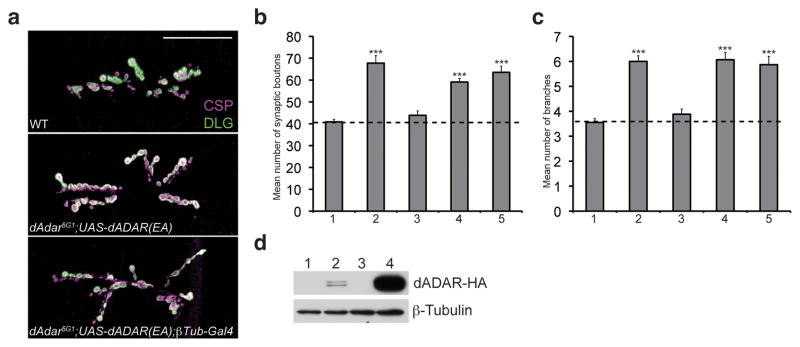
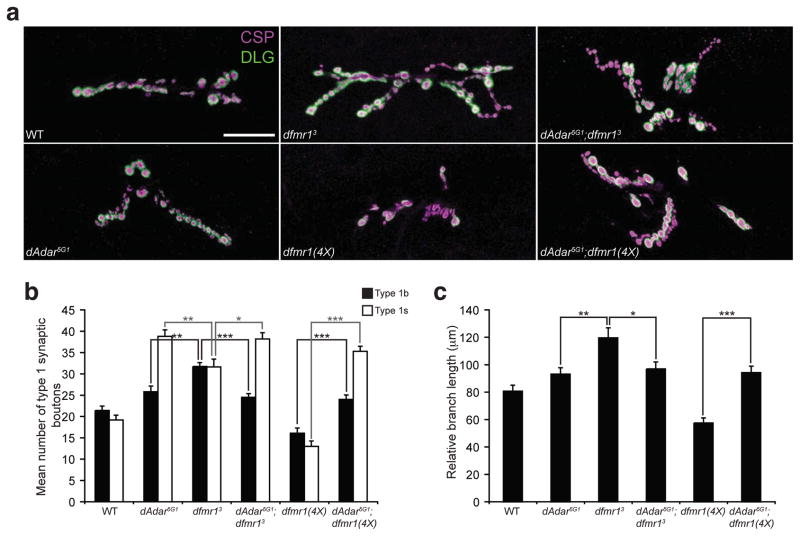
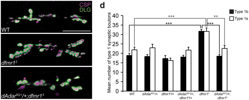
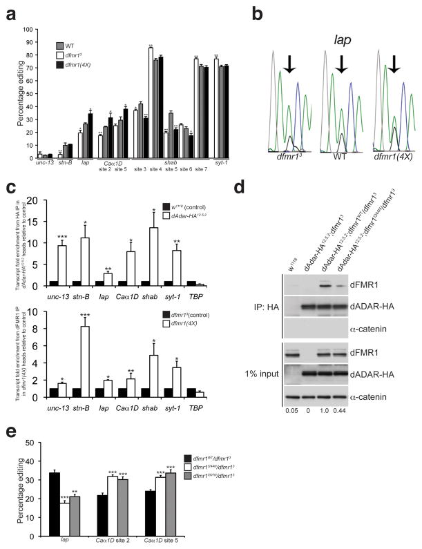
Loss of FMR1 gene function results in fragile X syndrome, the most common heritable form of intellectual disability. The protein encoded by this locus (FMRP) is an RNA-binding protein that is thought to primarily act as a translational regulator; however, recent studies have implicated FMRP in other mechanisms of gene regulation. We found that the Drosophila fragile X homolog (dFMR1) biochemically interacted with the adenosine-to-inosine RNA-editing enzyme dADAR. Adar and Fmr1 mutant larvae exhibited distinct morphological neuromuscular junction (NMJ) defects. Epistasis experiments based on these phenotypic differences revealed that Adar acts downstream of Fmr1 and that dFMR1 modulates dADAR activity. Furthermore, sequence analyses revealed that a loss or overexpression of dFMR1 affects editing efficiency on certain dADAR targets with defined roles in synaptic transmission. These results link dFMR1 with the RNA-editing pathway and suggest that proper NMJ synaptic architecture requires modulation of dADAR activity by dFMR1.


Conflict of interest statement

Competing Interests Statement

The authors declare no financial conflict of interest.

Figures

Figure 1
Figure 1. dFMR1 biochemically interacts with dADAR in Drosophila S2 cell culture and in vivo
(a) Structure of TAP (consisting of 2X FLAG and protein A sequences separated by a TEV cleavage site), dADAR(3A)-TAP and dADAR(3/4)-TAP constructs used to generate stable S2 cell lines. Constructs are under control of an inducible metallothionein (MT) promoter. (b) Western analysis showing expression of constructs in transfected S2 cells. Untransfected S2 cells were used as a negative control for the FLAG antibody. Astericks denote non-specific bands present in all samples that were detected by the anti-FLAG antibody. Molecular weight (MW) on left is measured in kilodaltons (kDa). (c) Eluates from TAP pulldown followed by TEV cleavage show that dFMR1 associates with dADAR-TAP in the presence of RNase A. Samples treated or untreated with RNase A are designated as (+) or (−), respectively. α-catenin was used as a loading control and does not associate with dADAR-TAP. A FLAG antibody was used to detect TAP constructs in input lanes. (d) RT-PCR analysis (upper panel) and ethidium bromide staining of total RNA (lower panel) on RNase A-treated and control lysates, showing efficient RNA degradation in RNase-treated samples. For RT-PCR analysis (upper panel), samples treated or untreated with RNase A are designated as (+) or (−), respectively. Primers against ribosomal protein 49 (rp49) and α-Tubulin84D-Tub84D) were used for PCR amplification. Molecular weight marker (MW) denotes size migration in basepairs (bp). For ethidium bromide staining of total RNA (lower panel), total RNA from TAP, dADAR(3A)-TAP, and dADAR(3/4)-TAP lysates were treated with DNase I (D), RNase A (R) or were untreated (C). (e) Co-IP experiments performed on head lysates prepared from w1118 (control) and two independent endogenously tagged dADAR-HA fly lines, dAdar-HA4.5.2 and dAdar-HA12.5.2. An HA antibody was used to detect dADAR-HA and α-catenin was used as a loading control and negative control for the co-IP.
Figure 2
Figure 2. dAdar5G1 mutants exhibit NMJ defects in third instar larvae
(a,b) Confocal images of muscles 6/7 (a) or muscle 4 (b) from WT (w1118) and dAdar5G1 L3 larvae. Presynaptic vesicles were stained for Cysteine-string protein (CSP, magenta) and Discs-large (DLG, green) was used as a postsynaptic marker. Type 1b and 1s boutons were distinguished by 1) the intensity of DLG staining, as DLG fluorescence is more intense on type 1b boutons relative to type 1s boutons, and 2) size, as type 1b boutons are larger than type 1s boutons. DLG staining was observed on type 1s synaptic boutons in all genotypes, however images were taken to keep visualization of DLG on type 1s boutons low to distinguish between the bouton subclasses. No noticeable differences in CSP and DLG intensity were observed across genotypes. Arrows indicate type 1s synaptic boutons. Scale bar represents 50 μm. (c,d) Quantification of average number of type 1b, type 1s, and total type 1 synaptic boutons in muscles 6/7 (c) or muscle 4 (d) of WT (black bars) or dAdar5G1 null (white bars) larvae. All images and quantification were performed using abdominal hemisegment A3. n≥16 for each genotype. Error bars denote s.e.m. *p<0.05, ***p<0.0001, WT vs. dAdar5G1 by Student’s t-test.
Figure 3
Figure 3. Neuronal expression of dADAR is sufficient for normal NMJ synaptic architecture
(a) Confocal images of the NMJ from the following genotypes: WT, dAdar5G1 larvae carrying a wild-type UAS-dADAR transgene (dAdar5G1;UAS-dADAR), dAdar5G1;UAS-dADAR;βTub-Gal4 (ubiquitous expression), dAdar5G1;UAS-dADAR;elav-Gal4 (neuronal expression), dAdar5G1;UAS-dADAR;scratch-Gal4 (neuronal expression), dAdar5G1;UAS-dADAR;MHC-Gal4 (muscle expression), and dAdar5G1;UAS-dADAR/G14-Gal4 (muscle expression). Larvae were stained for CSP (presynaptic, magenta) and DLG (postsynaptic, green). No noticeable differences in CSP and DLG intensity were observed across genotypes. Scale bar represents 50 μm. (b) Western analysis on L3 brain extracts from the following genotypes: w1118 (negative control for the HA antibody), dAdar-HA12.5.2 (for endogenous levels), dAdar5G1;UAS-dADAR and dAdar5G1;UAS-dADAR;βTub-Gal4. An antibody against HA was used to detect dADAR-HA expression (upper panel). β-Tubulin (lower panel) was used as a loading control. (c,d) Quantification of the average number of type 1 synaptic boutons (c) and synaptic branching (d) in muscles 6/7 for all genotypes. All images and quantification were performed using abdominal hemisegment A3, muscles 6/7. n≥16 for each genotype. Error bars denote s.e.m. ***p<0.001 analyzed by one-way ANOVA, p<0.0001 overall, Tukey-Kramer post-test. See Supplemental Table 1 for control genotypes.
Figure 4
Figure 4. Deaminase activity by dADAR is essential for normal NMJ synaptic architecture
(a) Confocal images of the NMJ from the following genotypes: WT, dAdar5G1 larvae carrying a UAS-dADAR(EA) transgene to express a catalytic mutant form of dADAR (dAdar5G1;UAS-dADAR(EA)), and dAdar5G1;UAS-dADAR(EA);βTub-Gal4. Larvae were stained for CSP (presynaptic, magenta) and DLG (postsynaptic, green). No noticeable differences in CSP and DLG intensity were observed across genotypes. Scale bar represents 50 μm. (b,c) Quantification of the average number of type 1 synaptic boutons (b) and synaptic branches (c) in muscles 6/7 for WT (1), dAdar5G1;UAS-dADAR(WT) (2), dAdar5G1;UAS-dADAR(WT);βTub-Gal4 (3), dAdar5G1;UAS-dADAR(EA) (4), and dAdar5G1;UAS-dADAR(EA);βTub-Gal4 (5). All images and quantification were performed using abdominal hemisegment A3, muscles 6/7. n≥16 for each genotype. Error bars denote s.e.m. ***p<0.001 analyzed by one-way ANOVA, p<0.0001 overall, Tukey-Kramer post-test. (d) Western analysis on lysates purified from w1118 (1), dAdar-HA12.5.2 (2), dAdar5G1;UAS-dADAR(EA) (3), and dAdar5G1;UAS-dADAR(EA);βTub-Gal4 (4) L3 larval brains. As observed with the wild-type dADAR transgene (see Fig. 3b), dADAR(EA) expression was higher compared to endogenous dADAR levels. Upper panel shows dADAR-HA expression and β-Tubulin (lower panel) served as a loading control.
Figure 5
Figure 5. dAdar and dfmr1 genetically interact
(a) Confocal images of L3 larval NMJs from the following genotypes: WT, dAdar5G1, dfmr13, dfmr1(4X), dAdar5G1;dfmr13 and dAdar5G1;dfmr1(4X). L3 larvae were stained for CSP (presynaptic, magenta) and DLG (postsynaptic, green). Type 1b and type 1s synaptic boutons were distinguished as described in Fig. 2. No noticeable differences in CSP and DLG intensity were observed across genotypes. Scale bar represents 50 μm. (b) Genetic studies demonstrating that dAdar is epistatic to dfmr1 with respect to synaptic bouton formation. Quantification of average number of type 1b (black bars) and type 1s (white bars) synaptic boutons for genotypes shown in (a). (c) Genetic studies demonstrating that dAdar is epistatic to dfmr1 with respect to primary branch length. Relative primary branch length was quantified for all genotypes shown in (a) using anti-HRP staining. The length of the primary branch length was normalized to the length of the abdominal hemisegment, and mean relative primary branch length was measured and plotted for each genotype. All images and quantification were performed using muscles 6/7, hemisegment A3. n≥16 for each genotype. Error bars denote s.e.m. *p<0.05, **p<0.01, ***p<0.001, analyzed by one-way ANOVA, p<0.0001 overall, Tukey-Kramer post-test.
Figure 6
Figure 6. Reduction of dAdar dosage rescues the dfmr13 null NMJ defects in L3 larvae
(a–c) Confocal images of WT (a), dfmr13 (b), and dAdar5G1/+;dfmr13 (c) L3 larval NMJs. Larvae were stained for CSP (presynaptic, magenta) and DLG (postsynaptic, green). Type 1b and type 1s synaptic boutons were distinguished as described in Fig. 2. No noticeable differences in CSP and DLG intensity were observed across genotypes. Scale bar represents 50 μm. (d) The reduction of dAdar dosage rescues the dfmr1 synaptic bouton phenotype, as revealed by quantification of average number of type 1b (black bars) and type 1s (white bars) synaptic boutons for trans-heterozygous genotypes using the dAdar5G1 and dfmr13 mutant alleles. All images and quantification were performed using muscles 6/7, hemisegment A3. n≥16 for each genotype. Error bars denote s.e.m. **p<0.01, ***p<0.001, analyzed by one-way ANOVA, p<0.0001 overall, Tukey-Kramer post-test.
Figure 7
Figure 7. dFMR1 modifies dADAR function and affects A-to-I editing efficiency
(a) Percentage of editing observed in samples from WT (gray bars), dfmr13 (white bars), and dfmr1(4X) (black bars) larvae was quantified and graphed for the following transcripts: unc-13, stoned-B (stn-B), lap, Caα1D, shab, and synaptotagmin-1 (syt-1). n = 3–7 individual RT-PCR reactions for each site. *p<0.05, **p<0.01, WT vs. dfmr13 and WT vs. dfmr1(4X) were analyzed with Mann Whitney-U test. Error bars denote s.e.m. (b) Representative electropherograms for the lap edited adenosine sequenced from WT, dfmr13, and dfmr1(4X) whole larval cDNA. Arrows point to edited sites analyzed. Green peak represents unedited (A) site, and black peak represents edited (G) site. (c) dADAR and dFMR1 associate with edited transcripts in vivo. RNA immunoprecipitation of transcripts associating with dADAR-HA or dFMR1 in adult head lysates. Fold enrichment of transcripts in dAdar-HA12.5.2 samples relative to w1118 was performed for the dADAR-HA RNA immunoprecipitation in the upper graph, and fold enrichment of transcripts in dfmr1(4X) overexpressing flies relative to dfmr13 null flies is shown for the dFMR1 RNA immunoprecipitation (lower graph). TBP was used as an unedited, non-specific transcript. Quantification of transcript enrichment was performed by using quantitative RT-PCR and fold enrichment was normalized to fold change of actin mRNA. Results represent four independent immunoprecipitation experiments for each HA and dFMR1 RNA IP and quantitative RT-PCR experiments was performed using three technical replicates. *p<0.05, **p<0.01, ***p<0.001, analyzed with Student’s t-test. Error bars denote s.e.m. (d) A mutation in the KH1 RNA binding domain of dFMR1 reduces the robustness of the dFMR1:dADAR biochemical interaction. co-IP experiments were performed with flies containing a wild-type dFMR1 construct (dfmr1WT) or a construct containing a point mutation in the dFMR1 KH1 domain (dfmr1I244N) that were crossed to the dAdar-HA12.5.2;dfmr13 fly line. w1118 flies served as a negative control for the HA antibody. An HA antibody was used to detect dADAR-HA and α-catenin was used as a loading control and negative control for the co-IP. Expression levels of dFMR1 in the IP samples were normalized to dFMR1 input levels and average fold change relative to dAdar12.5.2;dfmr1WT/dfmr13 is denoted below each lane. Experiment was performed three times. (e) Mutations in the dFMR1 KH domains affect editing of lap and Caα1D. Percentage of editing observed in dfmr1WT/dfmr13 (black bars), dfmr1I244N/dfmr13 (white bars), and dfmr1I307N/dfmr13 (gray bars) whole larvae. n = 3 8 individual RT-PCR reactions for each site. **p<0.01, ***p<0.001, dfmr1WT/dfmr13 vs. dfmr1I244N/dfmr13 and dfmr1WT/dfmr13 vs. dfmr11307N/dfmr13 were analyzed with Mann Whitney-U test. Error bars denote s.e.m.

Comment in

References

    1. O’Donnell WT, Warren ST. A decade of molecular studies of fragile X syndrome. Annu Rev Neurosci. 2002;25:315–338. - PubMed
    1. Feng Y, et al. FMRP associates with polyribosomes as an mRNP, and the I304N mutation of severe fragile X syndrome abolishes this association. Mol Cell. 1997;1:109–118. - PubMed
    1. Corbin F, et al. The fragile X mental retardation protein is associated with poly(A)+ mRNA in actively translating polyribosomes. Hum Mol Genet. 1997;6:1465–1472. - PubMed
    1. Khandjian EW, Corbin F, Woerly S, Rousseau F. The fragile X mental retardation protein is associated with ribosomes. Nat Genet. 1996;12:91–93. - PubMed
    1. Eberhart DE, Malter HE, Feng Y, Warren ST. The fragile X mental retardation protein is a ribonucleoprotein containing both nuclear localization and nuclear export signals. Hum Mol Genet. 1996;5:1083–1091. - PubMed

Publication types

MeSH terms