Review
. 2012 May;1822(5):639-49.
doi: 10.1016/j.bbadis.2011.10.011. Epub 2011 Oct 19.

Abnormal mitochondrial dynamics and synaptic degeneration as early events in Alzheimer's disease: implications to mitochondria-targeted antioxidant therapeutics

Affiliations
Review

Abnormal mitochondrial dynamics and synaptic degeneration as early events in Alzheimer's disease: implications to mitochondria-targeted antioxidant therapeutics

P Hemachandra Reddy et al. Biochim Biophys Acta. 2012 May.

Abstract

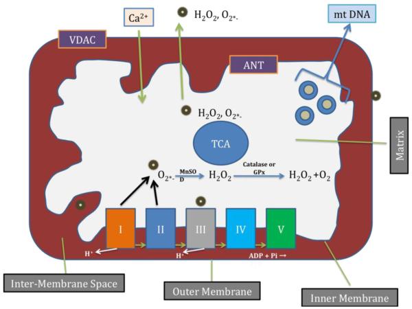
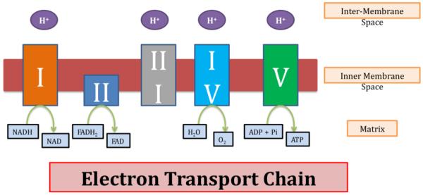
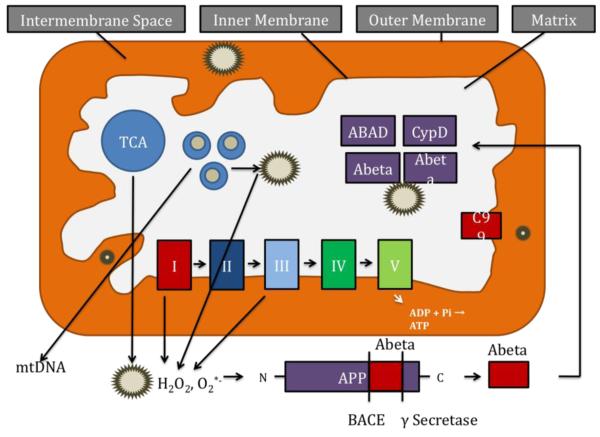
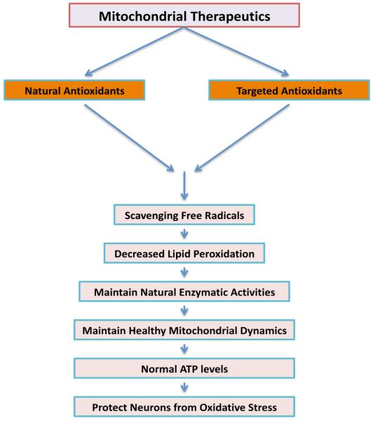
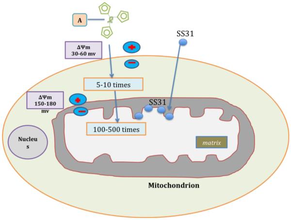
Synaptic pathology and mitochondrial oxidative damage are early events in Alzheimer's disease (AD) progression. Loss of synapses and synaptic damage are the best correlates of cognitive deficits found in AD patients. Recent research on amyloid beta (Aβ) and mitochondria in AD revealed that Aβ accumulates in synapses and synaptic mitochondria, leading to abnormal mitochondrial dynamics and synaptic degeneration in AD neurons. Further, recent studies using live-cell imaging and primary neurons from amyloid beta precursor protein (AβPP) transgenic mice revealed reduced mitochondrial mass, defective axonal transport of mitochondria and synaptic degeneration, indicating that Aβ is responsible for mitochondrial and synaptic deficiencies. Tremendous progress has been made in studying antioxidant approaches in mouse models of AD and clinical trials of AD patients. This article highlights the recent developments made in Aβ-induced abnormal mitochondrial dynamics, defective mitochondrial biogenesis, impaired axonal transport and synaptic deficiencies in AD. This article also focuses on mitochondrial approaches in treating AD, and also discusses latest research on mitochondria-targeted antioxidants in AD. This article is part of a Special Issue entitled: Antioxidants and Antioxidant Treatment in Disease.


Figures

Figure 1
Figure 1
Mitochondrial structure and sites of free radical generation. Mitochondria are bag like structures compartmentalized with two lipid membranes: the inner mitochondrial membrane and the outer mitochondrial membrane. The inner mitochondrial membrane houses the mitochondrial respiratory chain and provides a highly efficient barrier to ionic flow. The inner mitochondrial membrane houses respiratory chain or electron transport chain (ETC). In the ETC, complexes I and III leak electrons to oxygen, producing primarily superoxide radicals. Superoxide radicals are dismutated by manganese superoxide dismuase and produce H2O2. In addition, ETC involves H2O2 reducing to H2O and O2 by catalase or glutathione peroxidase accepting electrons donated by NADH and FADH2 and then yielding energy to generate ATP from adenosine diphosphate and inorganic phosphate. Free radicals are also generated by tricarboxylic acid in the matrix. These radicals are carried to the cytoplasm via voltage-dependent anion channels, and may involve oxidation DNA and proteins in the cytoplasm.
Figure 2
Figure 2
The structure of electron transport of chain.
Figure 3
Figure 3
Age and amyloid beta-induced free radical production and cleavage of APP fragments in AD neuron. The accumulation of mtDNA changes may induce ROS production and cause oxidative damage in aged tissues. In late-onset AD, age-dependent production of ROS contribute to the secretion of Aβ peptides by activating β- and γ-secretases. These Aβ peptides enter mitochondria, induce free radicals, decrease cytochrome oxidase activity, and inhibit ATP generation. In familial AD, mutations in APP, PS1 and PS2 activate β- and γ-secretases and secrete Aβ peptides, and these Aβ peptides enter mitochondria, cause mitochondrial dysfunction and damage neurons.
Figure 4
Figure 4
Schematic representation mitochondrial therapeutics for AD.
Figure 5
Figure 5
Schematic representation of targeting mitochondria by different molecules. A generic mitochondria-targeted antioxidant is shown constructed by the covalent attachment of an antioxidant molecule to the lipophilic triphenylphosphonium cation. Antioxidant molecules accumulate 5-10 fold in the cytoplasm, which is driven by plasma membrane potential, and then further accumulates 100-500 fold in the mitochondria. Mitochondria-targeted molecules rapidly neutralize free radicals and reduce mitochondrial toxicity. The SS31 is a cell-permeable tetra-peptide that targeted to mitochondria and protects mitochondria from oxidative damage. SS31 peptide has a sequence motif that allows them to target mitochondria several hundred fold more than natural antioxidants. Once SS peptides reach mitochondria, the SS peptides rapidly neutralize free radicals and decrease mitochondrial toxicity.

References

    1. Swerdlow RH. The neurodegenerative mitochondriopathies. J . Alzheimers. Dis. 2009;17:737–751. - PMC - PubMed
    1. Lin MT, Beal MF. Mitochondrial dysfunction and oxidative stress in neurodegenerative diseases. Nature. 2006;443:787–795. - PubMed
    1. Reddy PH, Beal MF. Are mitochondria critical in the pathogenesis of Alzheimer’s disease? Brain Res.Brain Res.Rev. 2005;49:618–632. - PubMed
    1. Reddy PH. Mitochondrial medicine for aging and neurodegenerative diseases. Neuromolecular Med. 2008;10:291–315. - PMC - PubMed
    1. Reddy PH. Role of mitochondria in neurodegenerative diseases: mitochondria as a therapeutic target in Alzheimer’s disease. CNS Spectr. 2009;14:8–13. discussion 16-8. - PMC - PubMed

Publication types

Substances