. 2012 Feb;1822(2):139-49.
doi: 10.1016/j.bbadis.2011.10.010. Epub 2011 Oct 20.

Compromised mitochondrial complex II in models of Machado-Joseph disease

Affiliations

Compromised mitochondrial complex II in models of Machado-Joseph disease

Mário N Laço et al. Biochim Biophys Acta. 2012 Feb.

Abstract

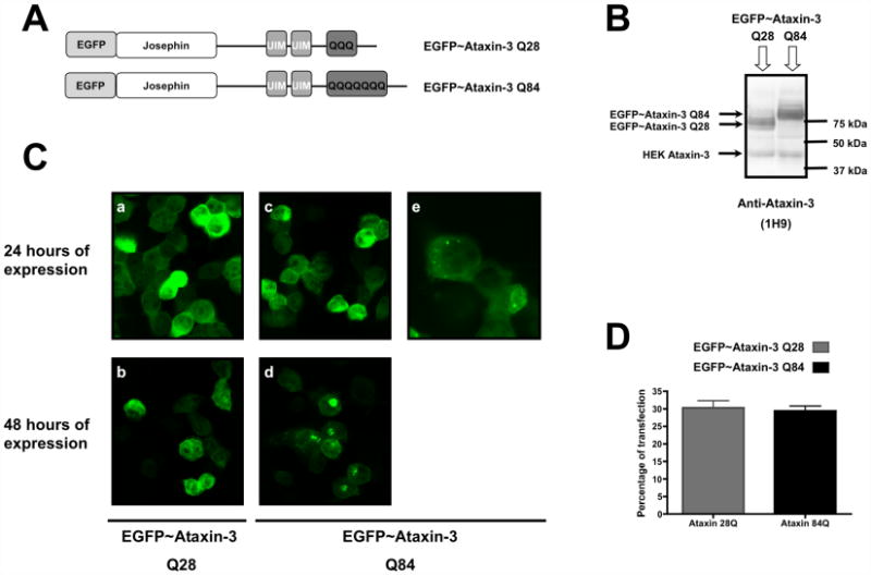
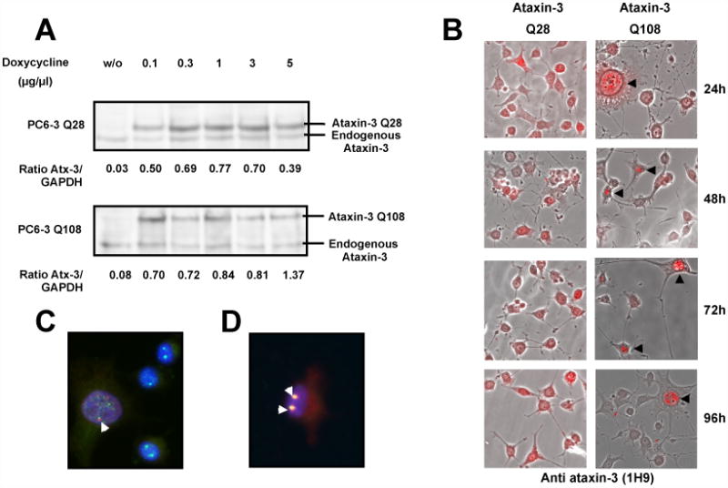
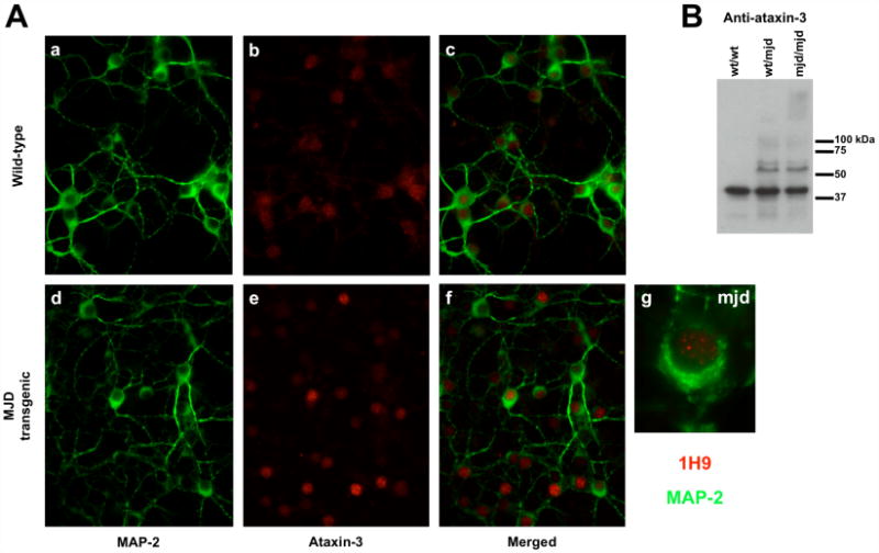
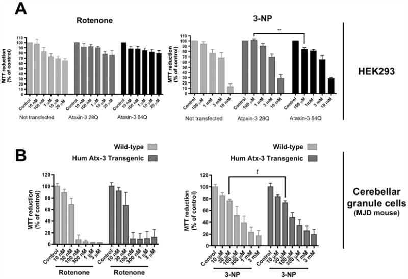
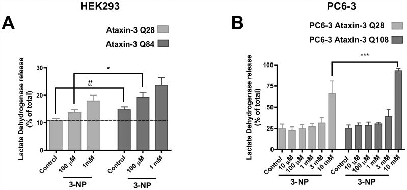
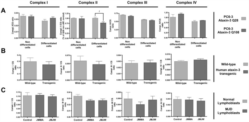
Machado-Joseph disease (MJD), also known as Spinocerebellar Ataxia type 3, is an inherited dominant autosomal neurodegenerative disorder. An expansion of Cytosine-Adenine-Guanine (CAG) repeats in the ATXN3 gene is translated as an expanded polyglutamine domain in the disease protein, ataxin-3. Selective neurodegeneration in MJD is evident in several subcortical brain regions including the cerebellum. Mitochondrial dysfunction has been proposed as a mechanism of neurodegeneration in polyglutamine disorders. In this study, we used different cell models and transgenic mice to assess the importance of mitochondria on cytotoxicity observed in MJD. Transiently transfected HEK cell lines with expanded (Q84) ataxin-3 exhibited a higher susceptibility to 3-nitropropionic acid (3-NP), an irreversible inhibitor of mitochondrial complex II. Increased susceptibility to 3-NP was also detected in stably transfected PC6-3 cells that inducibly express expanded (Q108) ataxin-3 in a tetracycline-regulated manner. Moreover, cerebellar granule cells from MJD transgenic mice were more sensitive to 3-NP inhibition than wild-type cerebellar neurons. PC6-3 (Q108) cells differentiated into a neuronal-like phenotype with nerve growth factor (NGF) exhibited a significant decrease in mitochondrial complex II activity. Mitochondria from MJD transgenic mouse model and lymphoblast cell lines derived from MJD patients also showed a trend toward reduced complex II activity. Our results suggest that mitochondrial complex II activity is moderately compromised in MJD, which may designate a common feature in polyglutamine toxicity.


Figures

Figure 1
Figure 1
EGFP-ataxin-3 Q84 fusion protein aggregates in a transiently expressed MJD cell model. HEK 293 cells were transfected with EGFP-ataxin-3 Q28 or EGFP-ataxin-3 Q84 plasmid constructs and their expression was carried out for 24 or 48 hours. (A) Representative schemes of the wild-type (Q28) and expanded (Q84) ataxin-3 fusion protein being expressed. (B) Total extracts prepared from HEK293 cells transfected with EGFP-ataxin-3 (Q28) or (Q84) were analysed through western blotting for ataxin-3 to assess the expression of the plasmid constructs. (C) Representative confocal fluorescent microscopy images of HEK293 cells expressing EGFP-ataxin-3 Q28 (a, b) or EGFP-ataxin-3 Q84 (c, d, e) for 24 (a, c, e) or 48 (b, d) hours. (e) Higher magnification of HEK cells transfected with EGFP-ataxin-3 Q84 for 24 hours. (D) HEK293 cells expressing EGFP-ataxin-3 Q28 or EGFP-ataxin-3 Q84 fusion proteins were fixed, nuclear stained with Hoechst 33342 and quantified through fluorescent microscopy. The graph plots the percentage of cells expressing wild-type (Q28) or expanded (Q84) ataxin-3 fluorescent fusion proteins in three independent transfections.
Figure 2
Figure 2
Aggregates of human expanded ataxin-3 in PC6-3 ataxin-3 Q108 cells are primarily nuclear. (A) Total extracts were prepared from PC6-3 ataxin-3 Q28 cells or PC6-3 ataxin-3 Q108 cells incubated in the absence or presence of increasing concentrations (0.1, 0.3, 1, 3 and 5 μg/μl) of doxycycline for 24 hours and subsequently probed for ataxin-3 on a western blot. (B) Merged representative images of fluorescent and optical differential interference contrast microscopy of PC6-3 cells incubated with doxycycline (1 μg/μl) for expression of human ataxin-3 Q28 or human ataxin-3 Q108 during 24, 48, 72 or 96 hours and immunostained for ataxin-3 (in red). (C, D) Representative images of PC6-3 ataxin-3 Q108 cells expressing human expanded ataxin-3 for 48 hours, which were fixed and immunostained for ataxin-3 (in red) and coilin (in green) (C) or PML protein (in green) (D). Yellow shows co-localization between proteins. Nuclei were stained with Hoechst 33342 (1 μg/ml).
Figure 3
Figure 3
Aggregation in the human expanded ataxin-3 transgenic mouse. (A) Cerebellar granule cells were isolated from 7 days wild-type (a, b, c) or transgenic (d, e, f) pups and kept in culture for 7 days. Cells were stained for MAP-2 (a, c, d, f - in green) and ataxin-3 (b, c, e, f - in red) and visualised through fluorescence microscopy. (g) Higher magnification of transgenic cerebellar granule cell immunostained for MAP-2 (in green) and ataxin-3 (in red). (B) Western blotting for ataxin-3 was performed in total brain extracts of wild-type (wt/wt), or transgenic (one copy (wt/mjd) or two copies (mjd/mjd) of the transgene) 4 month old mice to evaluate the expression of the transgene and the aggregation of human expanded ataxin-3.
Figure 4
Figure 4
Overexpression of expanded ataxin-3 (Q84) increases cell susceptibility to low concentrations of 3-nitropropionic acid (3-NP). HEK 293 cells transfected with EGFP-ataxin-3 (Q28) or (Q84) (A) or cerebellar granule cells from wild-type or transgenic mice pups (B) were incubated with rotenone (10 nM - 20 μM) or 3-NP (10 μM - 10 mM), for 24 hours. After this incubation period, cell viability was assessed by MTT (0.5 μg/ml) reduction. Graphs summarize the mean ± SEM of the percentage of control for each cell type of 4-8 independent experiments, run in duplicates. Statistical analysis: ** p<0.01, compared to HEK 293 EGFP-ataxin-3 Q28 treated with 100 μM 3-NP, two-way ANOVA followed by Bonferroni multiple comparison test; t p<0.05, compared to wild-type cerebellar granule cells treated with 30 μM 3-NP (Student's t-test).
Figure 5
Figure 5
Expanded ataxin-3 induces higher levels of cell death. HEK 293 cells expressing EGFP-ataxin-3 Q28 or Q84 (A) and PC6-3 cells expressing wild-type (Q28) or expanded (Q108) human ataxin-3 (B) were exposed to increasing concentrations of 3-NP (100 μM and 1 mM for HEK cells (A); 10 μM - 10 mM for PC6-3 cells (B)), for 24 hours. Total extracts and the extracellular media of every sample were collected and analysed for LDH activity to evaluate the degree of cell death. Graphs plot the mean ± SEM of the percentage of extracellular LDH activity of the total LDH activity, for each sample. 4-15 independent experiments were conducted. Statistical analysis: (A) tt p<0.01 compared to HEK 293 EGFP-ataxin-3 Q28 (Student's t-test); * p<0.05 compared to HEK 293 EGFP-ataxin-3 Q28 treated with 100 μM 3-NP, two-way ANOVA followed by Bonferroni multiple comparison test. (B) *** p<0.001 compared to PC6-3 Ataxin-3 Q28 treated with 10 mM 3-NP, two-way ANOVA followed by Bonferroni multiple comparison test.
Figure 6
Figure 6
Expanded ataxin-3 expression impairs mitochondrial complex II activity. The activities of complexes I, II, III and IV of the mitochondrial respiratory chain were determined spectrophotometrically in (A) mitochondrial extracts from undifferentiated and NGF-differentiated PC6-3 ataxin-3 Q28 and Q108 cell lines, (B) isolated brain mitochondria of 4-5 months wild-type or MJD homozygous transgenic mice, and (C) human lymphoblastic cell lines derived from MJD patients (JMMA, JMJW) or control individuals. Graph shows the mean ± SEM of 4-9 independent measurements of the mitochondrial complexes activities. The activities of mitochondrial complexes were normalised for citrate synthase activity. Statistical analysis: t p<0.05, compared to NGF-differentiated PC6-3 Ataxin-3 Q28 (Student's t-test).

References

    1. Albrecht M, Hoffman D, Evert BO, Scmitt I, Wullner U, Lengauer T. Structural modelling of ataxin-3 reveals distant homology to adaptins. Proteins. 2003;50:355–370. - PubMed
    1. Rüb U, Brunt ER, Del Turco D, de Vos RA, Gierga K, Paulson H, Braak H. The nucleus raphe interpositus in spinocerebellar ataxia type 3 (Machado-Joseph disease) J Chem Neuroanat. 2003;25:115–127. - PubMed
    1. Rüb U, de Vos RA, Schultz C, Brunt ER, Paulson H, Braak H. Spinocerebellar ataxia type 3 (Machado-Joseph disease): severe destruction of the lateral reticular nucleus. Brain. 2002;125:2115–2124. - PubMed
    1. Kawagushi Y, Okamoto T, Taniwaki M, Aizawa M, Inoue M, Katayama S, Kawakami H, Nakamura S, Nishimura M, Akiguchi I, et al. CAG expansions in a novel gene for Machado-Joseph disease at chromosome 14q32.1. Nat Genet. 1994;8:221–8. - PubMed
    1. Riess O, Rub U, Pastore A, Bauer P, Schols L. SCA3: Neurological features, pathogenesis and animal models. Cerebellum. 2008;7:125–137. - PubMed

Publication types

MeSH terms

LinkOut - more resources