. 2011 Dec 21;11(24):4274-8.
doi: 10.1039/c1lc20758j. Epub 2011 Oct 28.

Two-dimensional paper networks: programmable fluidic disconnects for multi-step processes in shaped paper

Affiliations

Two-dimensional paper networks: programmable fluidic disconnects for multi-step processes in shaped paper

Barry R Lutz et al. Lab Chip. .

Abstract

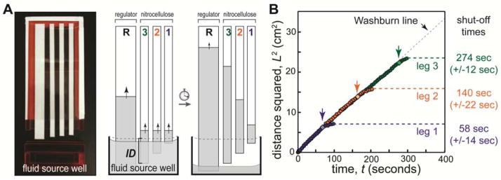
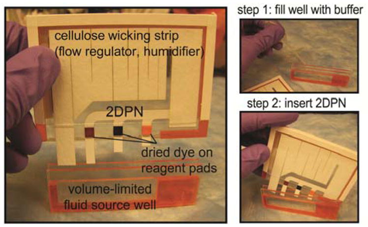
Most laboratory assays take advantage of multi-step protocols to achieve high performance, but conventional paper-based tests (e.g., lateral flow tests) are generally limited to assays that can be carried out in a single fluidic step. We have developed two-dimensional paper networks (2DPNs) that use materials from lateral flow tests but reconfigure them to enable programming of multi-step reagent delivery sequences. The 2DPN uses multiple converging fluid inlets to control the arrival time of each fluid to a detection zone or reaction zone, and it requires a method to disconnect each fluid source in a corresponding timed sequence. Here, we present a method that allows programmed disconnection of fluid sources required for multi-step delivery. A 2DPN with legs of different lengths is inserted into a shared buffer well, and the dropping fluid surface disconnects each leg at in a programmable sequence. This approach could enable multi-step laboratory assays to be converted into simple point-of-care devices that have high performance yet remain easy to use.


Figures

Figure 1
Figure 1
Volume-limited well for programmed fluid shut off. A) Nitrocellulose legs (width 5 mm) were arranged on an adhesive plastic backing (cartridge) such that each leg protruded a different length. An optional high capacity cellulose regulating wick (width 1 cm) was added to increase the rate of fluid depletion from the well and to humidify the device. The source well (1 cm tall, 5 cm wide, 2.5 mm thick) was filled with buffer (PBST), and the cartridge was inserted to initiate flow. The cartridge interior thickness (air gap) was 1.5 mm. The leg immersion depth (ID) is indicated for leg 3; immersion depths were 3.5 mm, 4.5 mm, 5.5 mm for legs 1, 2, and 3, respectively. As fluid was wicked, each leg broke contact with the fluid in a timed sequence to provide programmed shut off. B) Plot of the fluid front position in each leg as a function of time. The fluid front in each leg followed Lucas-Washburn (L2=Wt) until it broke contact with the fluid in the well (indicated by light diamonds and arrows). The fluid front migrated somewhat after disconnection due to relaxation of fluid into the dry membrane. Relative errors in shut off times were 20%, 15%, and 5% for legs 1, 2 and 3, respectively.
Figure 2
Figure 2
A complete 2DPN for autonomous sequential fluid delivery. A) The 2DPN includes mock reagents (dyes) dried on pads and a regulating wick in a plastic housing. The cellulose wick regulated the fluid depletion rate, served as the downstream wicking pad, and humidified the device. Nitrocellulose legs: 0.5 cm wide, horizontal spacing 1 cm; leg immersion depths 4 mm (yellow), 7 mm (blue), and 13 mm (red). Regulating wick: cellulose, 1 cm wide at base. Reagent pads: 0.5cm x 0.5 cm cellulose with dried food colouring. Well dimensions: 6 cm wide x 1.5 cm tall x 2.5 mm thick; cartridge interior thickness (air gap): 1 mm. B) The device is operated by filling the self-metering well to capacity and inserting the 2DPN cartridge to initiate the sequence. The cartridge was designed to be fully inserted against a mechanical stop in the well, such that careful positioning by the user was not required.
Figure 3
Figure 3
Autonomous sequential fluid delivery in a 2DPN. Each leg wicked fluid from a single buffer source, and dried dyes representing reagents create different fluids from each leg (colours). Each coloured fluid arrived at a different time at the “detection zone” (green box) and was shut off in a timed sequence after delivery. Some “bleeding” from the cellulose reagent pads can be seen as a narrow band that occupies about 20% of the strip width. Time courses show colour across the full strip width or the detection zone as a function of time after device activation. Device dimensions are given in Figure 2. For perspective, the 2DPN shown here is ~2.5-fold larger in area than a conventional LFT (e.g., LFT: 5mm x 40mm = 200mm2; 2DPN paper area: 500mm2), but smaller devices (2DPN and well) can be made if desired.

References

    1. Pelton R. Bioactive paper provides a low-cost platform for diagnostics. Trac-Trends Anal Chem. 2009;28(8):925–942. - PMC - PubMed
    1. Zhao WA, et al. Paper-Based Bioassays Using Gold Nanoparticle Colorimetric Probes. Anal Chem. 2008;80(22):8431–8437. - PubMed
    1. Dungchai W, et al. Electrochemical Detection for Paper-Based Microfluidics. Anal Chem. 2009;81(14):5821–5826. - PubMed
    1. Carvalhal RF, et al. Electrochemical Detection in a Paper-Based Separation Device. Anal Chem. 2010;82(3):1162–1165. - PubMed
    1. Nie ZH, et al. Electrochemical sensing in paper-based microfluidic devices. Lab Chip. 2010;10(4):477–483. - PMC - PubMed

Publication types