

Review
doi: 10.1016/j.gene.2011.10.030.
Epub 2011 Oct 21.
Biochemistry and biology of the inducible multifunctional transcription factor TFII-I: 10 years later
Affiliations
- PMID: 22037610
- PMCID: PMC3246126
- DOI: 10.1016/j.gene.2011.10.030
Item in Clipboard
Review
Biochemistry and biology of the inducible multifunctional transcription factor TFII-I: 10 years later
Gene.
.
Abstract
Exactly twenty years ago TFII-I was discovered as a biochemical entity that was able to bind to and function via a core promoter element called the Initiator (Inr). Since then several different properties of this signal-induced multifunctional factor were discovered. Here I update these ever expanding functions of TFII-I--focusing primarily on the last ten years since the first review appeared in this journal.
Copyright © 2011 Elsevier B.V. All rights reserved.
Figures
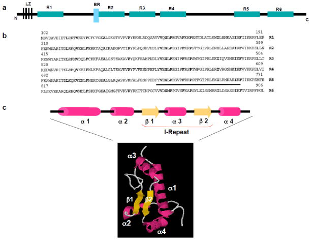
a) Schematic of TFII-I with 6 repeated regions, R1–R6, the N-terminal leucine zipper (LZ) and the basic region (BR) which serves as a DNA binding domain. B) The sequence of the repeats R1–R6. The most conserved amino acids are indicated in bold and the I-repeat is underlined. c) Both the linear arrangement and NMR structure of the murine repeat 5 (R5). The various amphipathic helices are indicated as α1–4 and the beta sheets are indicated as β1 and β2. The figure depicting the NMR structure is kindly provided by H. Hirota. This unique structural fold is referred to as an I-fold.
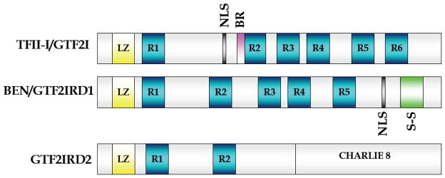
TFII-I, also known as GTF2I has 6 repeats (R1–R6) with the NLS and BR preceding R2. BEN (GTF2IRD1/MusTRD1/GTF3) has five repeats with the NLS located toward the C-terminal region. The S-S is a serine stretch, the precise function of which is unknown. The third member, GTF2IRD2 has only two repeats (R1 and R2) and does not have any NLS. The N-terminal region of this member shares 75% identity with TFII-I and resembles a truncated TFII-I. GTF2IRD2 has a Charlie8 transposon-like domain. However, the function of this domain is currently unknown. The N-terminal leucine zipper (LZ) is well conserved amongst the three members.
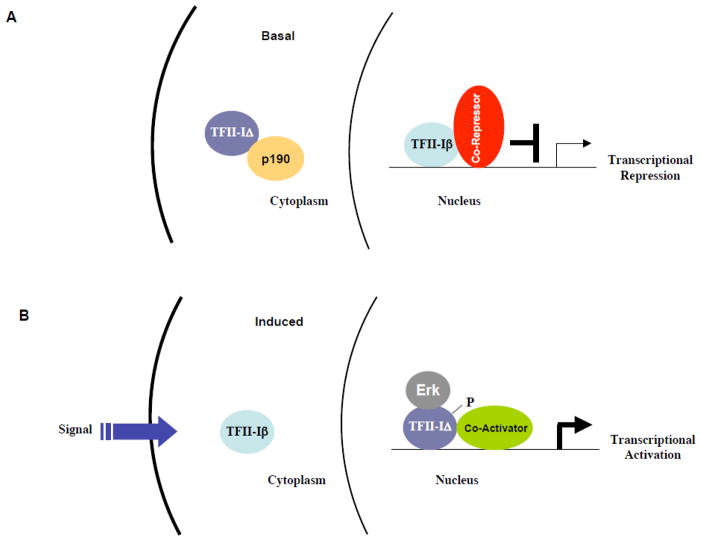
(A) In basal state, unphosphorylated TFII-IΔ is predominantly cytoplasmic, tethered by p190RhoGAP (p190). Other molecules can also tether TFII-I in the cytoplasm (not shown for simplicity). TFII-Iβ is recruited to a c-fos promoter site, likely in conjunction with co-repressors (red) (e.g., HDAC1 and 3, LSD1 or PRC) resulting in a transcriptional repression. (B) Upon signaling, TFII-IΔ undergoes phosphorylation, interacts with Erk1/2 and translocate to the nucleus. Under these conditions, TFII-Iβ is exported out of the nucleus and instead TFII-IΔ occupies the same site on the c-fos promoter, resulting in transcriptional activation. It is conjectured that under these conditions, TFII-IΔ interacts with transcriptional co-activators (green), although the precise identities of these factors are currently unknown.
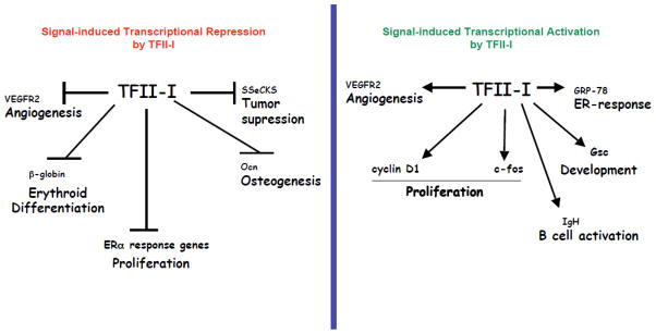
Shown on the left, signal -dependent transcriptional repression mediated by TFII-I (in red). The target genes and functional consequences are indicated. Shown on the right, signal-dependent transcriptional activation mediated by TFII-I (in green). These functions are likely to be signal and cell type dependent as activation and repression of both VEGFR2 and cyclin D1 have been shown.
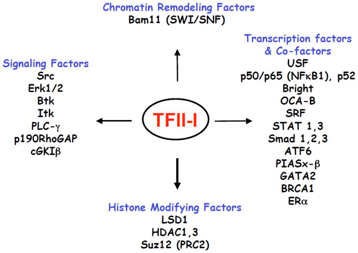
The interacting proteins are divided into four functional groups: transcription factors/co-factors, chromatin modifying factors, histone modifying factors and signaling molecules. These interactions are observed in different cell types and under distinct conditions.
Similar articles
-
Functional analysis of TFIID components.Cold Spring Harb Symp Quant Biol. 1998;63:219-27. doi: 10.1101/sqb.1998.63.219. Cold Spring Harb Symp Quant Biol. 1998. PMID: 10384285 Review. No abstract available.
-
Williams-Beuren syndrome-associated transcription factor TFII-I regulates osteogenic marker genes.J Biol Chem. 2009 Dec 25;284(52):36234-36239. doi: 10.1074/jbc.C109.063115. Epub 2009 Oct 30. J Biol Chem. 2009. PMID: 19880526 Free PMC article.
-
Functional interrelationship between TFII-I and E2F transcription factors at specific cell cycle gene loci.J Cell Biochem. 2018 Jan;119(1):712-722. doi: 10.1002/jcb.26235. Epub 2017 Jul 31. J Cell Biochem. 2018. PMID: 28657656 Free PMC article.
-
A Novel Interaction between TFII-I and Mdm2 with a Negative Effect on TFII-I Transcriptional Activity.PLoS One. 2015 Dec 11;10(12):e0144753. doi: 10.1371/journal.pone.0144753. eCollection 2015. PLoS One. 2015. PMID: 26656605 Free PMC article.
-
TFII-IDelta and TFII-Ibeta: unequal brothers fostering cellular proliferation.Mol Cell. 2006 Oct 20;24(2):169-71. doi: 10.1016/j.molcel.2006.10.003. Mol Cell. 2006. PMID: 17052451 Review.
Cited by
-
Deletion of Gtf2i via Systemic Administration of AAV-PHP.eB Virus Increases Social Behavior in a Mouse Model of a Neurodevelopmental Disorder.Biomedicines. 2023 Aug 15;11(8):2273. doi: 10.3390/biomedicines11082273. Biomedicines. 2023. PMID: 37626769 Free PMC article.
-
The transcription factor TFII-I promotes DNA translesion synthesis and genomic stability.PLoS Genet. 2014 Jun 12;10(6):e1004419. doi: 10.1371/journal.pgen.1004419. eCollection 2014 Jun. PLoS Genet. 2014. PMID: 24922507 Free PMC article.
-
Cellular transcription factor TFII-I represses adenovirus gene expression.J Virol. 2025 Jun 17;99(6):e0061825. doi: 10.1128/jvi.00618-25. Epub 2025 May 12. J Virol. 2025. PMID: 40353670 Free PMC article.
-
Autographa californica multiple nucleopolyhedrovirus orf114 is not essential for virus replication in vitro, but its knockout reduces per os infectivity in vivo.Virus Genes. 2012 Oct;45(2):360-9. doi: 10.1007/s11262-012-0777-y. Epub 2012 Jun 28. Virus Genes. 2012. PMID: 22739701
-
TFII-I/Gtf2i and Erythro-Megakaryopoiesis.Front Physiol. 2020 Sep 25;11:590180. doi: 10.3389/fphys.2020.590180. eCollection 2020. Front Physiol. 2020. PMID: 33101065 Free PMC article.
References
-
- Abdelrahim M, Liu S, Safe S. Induction of endoplasmic reticulum-induced stress genes in Panc-1 pancreatic cancer cells is dependent on Sp proteins. J Biol Chem. 2005;280:16508–16513. - PubMed
-
- Ashworth T, Roy AL. Phase specific functions of the transcription factor TFII-I during cell cycle. Cell Cycle. 2009;8:596–605. - PubMed
-
- Ashworth T, Roy AL. Cutting Edge: TFII-I controls B cell proliferation via regulating NF-kappaB. J Immunol. 2007;178:2631–2635. - PubMed
Publication types
MeSH terms
Substances
Grants and funding
LinkOut - more resources
Full Text Sources
Other Literature Sources
