

Cardiomyopathy in the mouse model of Duchenne muscular dystrophy caused by disordered secretion of vascular endothelial growth factor
- PMID: 22037736
- PMCID: PMC3539494
- DOI: 10.12659/msm.882043
Cardiomyopathy in the mouse model of Duchenne muscular dystrophy caused by disordered secretion of vascular endothelial growth factor
Abstract
Background: Duchenne muscular dystrophy (DMD) is a genetic neuromuscular disorder that affects skeletal muscles and cardiac muscle tissue. In some cases, myocardial injury secondary to hypoxia can lead to dilative cardiomyopathy (DCM). A genetic defect in the dystrophin gene may increase the susceptibility of myocardium to hypoxia. Available data suggest that this may be caused by impaired secretion of NO, which is bound with secretion of VEGF-A.
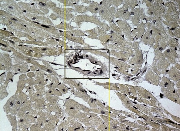
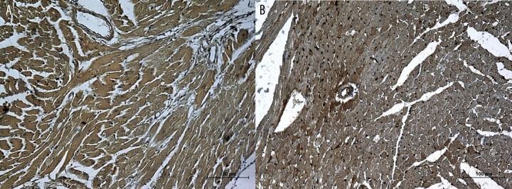
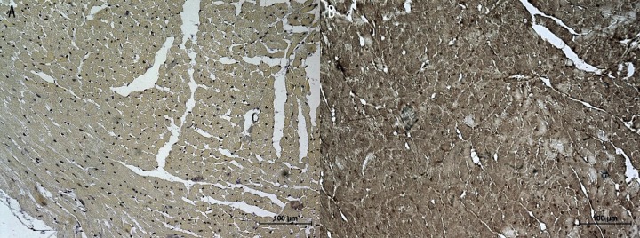
Material/methods: Male mice C57BI/10ScSn mdx (animal model of DMD) and healthy mice C57BI/10ScSn were exposed to hypobaric hypoxia in low-pressure chambers. Their hearts were harvested immediately after and 1, 3, 7, and 21 days after exposure to hypoxia. Normobaric mice were used as controls. The expression of VEGF-A in myocardium and cardiac vessel walls was evaluated using immunohistochemistry, Western blotting, and in situ hybridization.
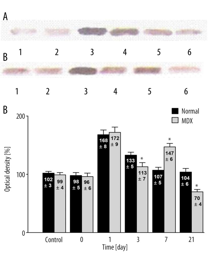
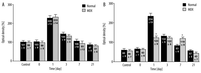
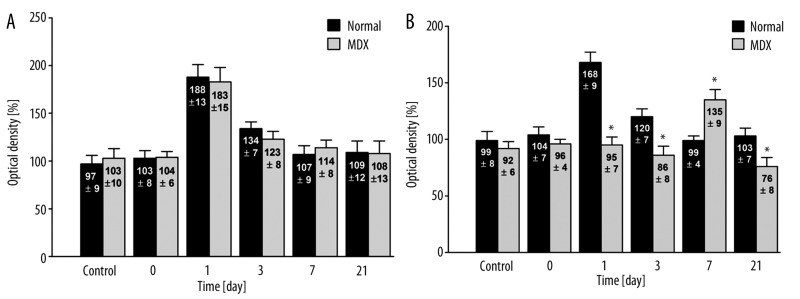
Results: VEGF-A expression in myocardium and vessel walls of healthy mice peaked 24 hours after exposure to hypoxia. The expression of VEGF-A in vessel walls was similar in dystrophic and healthy mice; however, VEGF-A expression in the myocardium of dystrophic mice was impaired, peaking around day 7. In the heart, the total level of VEGF depends on VEGF expression in myocardium, not in vessel endothelium, and our research demonstrates that the expression of VEGF is dystrophin-dependent.
Conclusions: Disordered secretion of VEGF-A in hypoxic myocardium caused the total level of this factor to be impaired in the heart. This factor, which in normal situations protect against hypoxia, promotes the gradual progression of cardiomyopathy.
Figures
References
-
- Emery AE. Population frequencies of inherited neuromuscular diseases-a world survey. Neuromuscul Disord. 1991;1(1):19–29. - PubMed
-
- Baxter P. Treatment of the heart in Duchenne muscular dystrophy. Dev Med Child Neurol. 2006;48(3):163. - PubMed
-
- de Kermadec JM, Becane HM, Chenard A, et al. Prevalence of left ventricular systolic dysfunction in Duchenne muscular dystrophy: an echocardiographic study. Am Heart J. 1994;127(3):618–23. - PubMed
-
- Eagle M, Baudouin SV, Chandler C, et al. Survival in Duchenne muscular dystrophy: improvements in life expectancy since 1967 and the impact of home nocturnal ventilation. Neuromuscul Disord. 2002;12(10):926–29. - PubMed
-
- Finsterer J, Stollberger C. The heart in human dystrophinopathies. Cardiology. 2003;99(1):1–19. - PubMed
Publication types
MeSH terms
Substances
LinkOut - more resources
Full Text Sources
Medical
