. 2011 Dec;11(24):4660-76.
doi: 10.1002/pmic.201100058. Epub 2011 Nov 23.

Proteomic analysis of Col11a1-associated protein complexes

Affiliations

Proteomic analysis of Col11a1-associated protein complexes

Raquel J Brown et al. Proteomics. 2011 Dec.

Abstract

Cartilage plays an essential role during skeletal development within the growth plate and in articular joint function. Interactions between the collagen fibrils and other extracellular matrix molecules maintain structural integrity of cartilage, orchestrate complex dynamic events during embryonic development, and help to regulate fibrillogenesis. To increase our understanding of these events, affinity chromatography and liquid chromatography/tandem mass spectrometry were used to identify proteins that interact with the collagen fibril surface via the amino terminal domain of collagen α1(XI) a protein domain that is displayed at the surface of heterotypic collagen fibrils of cartilage. Proteins extracted from fetal bovine cartilage using homogenization in high ionic strength buffer were selected based on affinity for the amino terminal noncollagenous domain of collagen α1(XI). MS was used to determine the amino acid sequence of tryptic fragments for protein identification. Extracellular matrix molecules and cellular proteins that were identified as interacting with the amino terminal domain of collagen α1(XI) directly or indirectly, included proteoglycans, collagens, and matricellular molecules, some of which also play a role in fibrillogenesis, while others are known to function in the maintenance of tissue integrity. Characterization of these molecular interactions will provide a more thorough understanding of how the extracellular matrix molecules of cartilage interact and what role collagen XI plays in the process of fibrillogenesis and maintenance of tissue integrity. Such information will aid tissue engineering and cartilage regeneration efforts to treat cartilage tissue damage and degeneration.


Conflict of interest statement

Authors have no declared financial or commercial conflicts of interest.

Figures

Figure 1
Figure 1
Schematic diagram of collagen fibrils with collagen type XI (blue) located within the interior of type II collagen fibrils (orange). The globular nature of collagen α1(XI) NTD leads to the restricted localization on the surface of collagen fibrils even though the triple helical domain of type XI collagen lies in the interior of the fibril. The retained NTD may sterically hinder the further addition of collagen molecules and thereby limit the ultimate diameter of the collagen fibril. Additionally, the NTD of collagen α1(XI) on the surface of the fibril may interact with other matrix components.
Figure 2
Figure 2
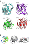
(A) Structural depictions of NTD of α1(XI) homology models (grey) aligned with respective templates [2UUR(blue), 21R6(green), 1LHN(red), and 1Z78(purple)]. (B) The α1(XI) NTD homology model from 2UUR (left), model with putative binding site (center) and zoom image of binding region (right).
Figure 3
Figure 3
SDS-PAGE of recombinant α1(XI) NTD, cartilage extract, and affinity selected proteins. Proteins were separated by SDS-PAGE and stained with Coomassie Safe Blue. (A) Molecular weight markers (lane 1), purified recombinant α1(XI) NTD protein (lane 2), proteins bound to nonderivatized column resin indicating nonspecific binding (lane 3), and total protein extract (lane 4). Bands or regions of the gel from lane 4 were manually excised, trypsinized, and analyzed by mass spectrometry to determine identity. For each region of the gel, the following proteins were identified: In the 240/230Mr range: biglycan, collagen α1(XI), actin, aggrecan, perlecan, collagen α1(XII), decorin, collagen α1(I), epiphycan, and collagen α1(XIV); in the 210Mr range: collagen α1(XIV), collagen α1(XII), biglycan, and tenascin-C; in the 180Mr range:thrombospondin-1, biglycan, collagen α1(XIV), perlecan, biglycan, collagen α1(XII), nidogen2,tenascin-C, collagen α1(I), collagen α1(XII), and fibromodulin; in the 160Mr range: collagen α1(II), biglycan, thrombospondin-1, fibromodulin, and collagen α1(I); in the 140Mr range: biglycan and COMP; in the 130Mr range: COMP and thrombospondin-1; in the 120Mr range: COMP, collagen α1(IX), biglycan, collagen α1(XIV), fibromodulin, thrombospondin-1, and matrilin-3; in the 100Mr range: biglycan, actinin alpha-4,fibromodulin, collagen α1(IX), matrilin-3, collagen α1(XIV), thrombospondin-1, collagen α1(II), nucleolin, pyruvate kinase, ovalbumin, and elongation-factor 2; in the 75Mr range: fibromodulin, lysyl hydroxylase, biglycan, collagen α1(II), thrombospondin-1, protein disulfide isomerase A4, and heat shock protein (GRP78); in the 60Mr range: CMP, protein disulfide isomerase A4, heat shock protein (GRP78), transferrin, semenogelin/inhibin, protein disulfide isomerase A1, actin, pyruvate kinase M2, collagens, phosphoglycerate kinase, vitrin, calreticulin, chondrocalcin, protein disulfide isomerase A5, and PARP; in the 48Mr range: actin, phosphoglycerate kinase, collagen-binding protein, and vitrin; in the 45Mr range: collagen α1(XI) NTD, fructose bisphosphate aldolase, CMP, and cartilage link protein; in the 40Mr range: chondroadherin, chondrocalcin, CMP, fructose bisphosphate aldolase, cartilage link protein, actin, and collagen α1(XI) NTD; in the 38Mr range: annexin A1, annexin A2, annexin A5, chondrocalcin, chondroadherin, lactate dehydrogenase B, lactate dehydrogenase A, collagen α1(XI) NTD, PARP, CMP, thrombosponin-1, fructose bisphosphate aldolase, and actin; in the 35Mr range: chondrocalcin, chondroadherin, annexin A1, annexin A2, annexin A5, CMP, thrombospondin-1, PARP, actin, lactate dehydrogenase A, and collagen α1(XI) NTD; in the 32Mr range: chondrocalcin, chondroadherin, annexin A1, annexin A2, annexin A5, thrombospondin-1, lactate dehydrogenase A, collagen α1(XI) NTD, and ANP32B; in the 28Mr range: collagen α1(I), PARP, chondrocalcin, CMP, thrombospondin-1, and collagen α1(XI) NTD, in the 25Mr range: PARP and CMP; in the 22Mr range: keratin; in the 20Mr range: lectin, histone 2A, histone 2B, collagen α1(XI) NTD; and in the 18Mr range: hemoglobin. (B). Cartilage proteins that interact with the NTD of collagen α1(XI). SDS-PAGE stained with Coomassie Safe Blue. Molecular weight markers (lane 1), proteins selected by affinity chromatography (lane 2). Bands or regions of the gel indicated by letters a through p on the right-hand side of gel were manually excised, trypsinized, and analyzed by mass spectrometry. Proteins identified in the specific locations a through p were as follows: a) (240Mr): biglycan; b) (180Mr) collagen α1(XIV), thrombospondin-1, perlecan, biglycan, and collagen α1(XII); c) (160Mr)collagen α1(XII), biglycan, thrombospondin-1, and fibromodulin; d) (130Mr) thrombospondin-1; e) (120Mr) COMP, collagen α1(IX), biglycan, collagen α1(XIV), fibromodulin, thrombospondin-1, and matrilin-3; f) (100Mr) biglycan, actinin alpha-4, fibromodulin, collagen α1(IX), PARP, matrilin-3, collagen α1(XIV),thrombospondin-1, and collagen α1(XII) ; g) (90Mr) nucleolin and fibromodulin; h) (75Mr) fibromodulin, lysyl hydroxylase 1, biglycan, collagen α1(XII), thrombospondin-1, protein disulfide A4, transferrin, and heat shock protein 70; i) (60Mr) CMP, protein disulfide A3, fibromodulin, chondrocalcin, PARP, and calreticulin; j) (45Mr) CMP; k) (40Mr) CMP and 1,6 fructose bisphosphate aldolase; l) (38Mr) chondrocalcin, annexin A2, annexin A1, annexin A5, CMP, PARP, thrombospondin-1, 1,6 fructose bisphosphate and actin; m) (36Mr) chondrocalcin, annexin A5, annexin A1, annexin A2, CMP. thrombospondin -1, PARP, and actin; n) (34Mr) chondrocalcin, acidic leucine-rich nuclear phosphoprotein 32 family member B, annexin A1, annexin A2, annexin A5, collagen α1(XI) NTD, chondroadherin, lactate dehydrogenase A, thrombospondin-1, PARP, and CMP; o) (29Mr) PARP, chondrocalcin, CMP, thrombospondin-1, 1,6- fructose bisphosphate aldolase, and actin; p) (27Mr): PARP, thrombospondin-1, and collagen α1(XI) NTD. (C) Grid depicting proteins identified as a function of location on gel. Apparent molecular weight is indicated on the vertical axis and protein identity is indicated along the horizontal axis.
Figure 4
Figure 4
Extracellular matrix network of predicted interactions. A subset of the identified proteins are shown as a network of interacting proteins based upon this study and evidence from previously published work. (A). Col2a1, CHAD, THBS1, MATN1, MATN3, and COMP are shown within the STRING network seeded by Col11a1, where confidence is indicated by the number of lines connecting the protein nodes of the network to each other and the color in the grid shown in (B). The color of the line is indicative of the type of data supporting the predicted interaction. Purple indicates that experimental or biochemical evidence for an interaction exists in the literature, blue indicates co-occurrence evidence, purple indicates experimental evidence, yellow indicates evidence from text-mining that detected co-mentioned in PubMed abstract, light blue indicates the association in curated database, grey indicates predicted interaction based on sequence/structural homology.
Figure 5
Figure 5
Extended network of predicted interactions. Interactions that include extracellular matrix, membrane and cellular proteins are shown to include predicted primary and secondary interactions. Each protein detected in the affinity experiment was analyzed using STRING molecular interactions. Thickness of connecting lines indicates confidence level associated with each interaction.
Figure 6
Figure 6
Interaction between thrombospondin 1 and the NTD of α1(XI) collagen was analyzed by surface plasmon resonance. Collagen α1(XI) NTD was covalently coupled to the sensor chip, while thrombospondin 1 at concentrations ranging from 15 to 500 nM was allowed to bind to the Collagen α1(XI) NTD. The average of 3 runs to collect association data is shown. Using the data shown to fit to a steady state affinity model, the dissociation constant (Kd) was calculated to be 100nM for native thrombospondin 1, assuming a one-site association model.
Figure 7
Figure 7
Colocalization of thrombospondin 1 and collagen type XI alpha 1 chain by immunofluorescence. A polyclonal antibody to the rat α1(XI) collagen α1(XI) and a monoclonal antibody to thrombospondin-1 were used to detect these ECM molecules within newly synthesized pericellular matrix from a cell line derived from the Swarm rat chondrosarcoma. Cells were plated at 3.5 × 10⁴ cells/cm2 onto poly-d-lysine/laminin-coated chamber slides and accumulated pericellular matrix was analyzed after 48 hours. Primary antibodies were detected with secondary antibody, rhodamine (TRITC)-conjugated AffiniPure donkey anti-rabbit IgG or fluorescein isothiocyanate-conjugated anti-mouse IgG and counterstained with DAPI. (A) Thrombospondin-1 antibody (green), (B) Collagen α1(XI) antibody (red), (C) Thrombospondin-1 antibody (green) and Collagen α1(XI) (red) overlap resulting in yellow where colocalization occurs.

Similar articles

Cited by

References

    1. Pogue R, Sebald E, King L, Kronstadt E, et al. A transcriptional profile of human fetal cartilage. Matrix Biology. 2004;23:299–307. - PubMed
    1. Poole CA. In: Joint Cartilage Degradation; Basic and Clinical Aspects. Woessner JF, Howell DS, editors. New York: Marcell Dekker; 1993.
    1. Wight TN, Heinegard DK, Hascall VC. In: Cell Biology of the Extracellular Matrix. Hay ED, editor. New York: Plenum; 1991. pp. 45–78.
    1. Hardingham TE, Venn G. Chondrotin sulphate/dermatan sulfate proteoglycans in cartilage: aggrecan, decorin and biglycan. In: Scott JE, editor. Dermatan Sulfate Proteoglycans: Chemistry, Biology, Chemical Pathology. London, England: Portans Press; 1993. pp. 207–217.
    1. Burgeson RE, Hollister DW. Collagen heterogeneity in human cartilage: identification of several new collagen chains. Biochem. Biophys. Res. Com. 1979;87:1124–1131. - PubMed

Publication types

Substances

LinkOut - more resources