. 2011 Dec 15;17(24):7595-604.
doi: 10.1158/1078-0432.CCR-11-1456. Epub 2011 Oct 28.

Targeted therapy for BRAFV600E malignant astrocytoma

Affiliations

Targeted therapy for BRAFV600E malignant astrocytoma

Theodore P Nicolaides et al. Clin Cancer Res. .

Abstract

Purpose: Malignant astrocytomas (MA) are aggressive central nervous system tumors with poor prognosis. Activating mutation of BRAF (BRAF(V600E)) has been reported in a subset of these tumors, especially in children. We have investigated the incidence of BRAF(V600E) in additional pediatric patient cohorts and examined the effects of BRAF blockade in preclinical models of BRAF(V600E) and wild-type BRAF MA.

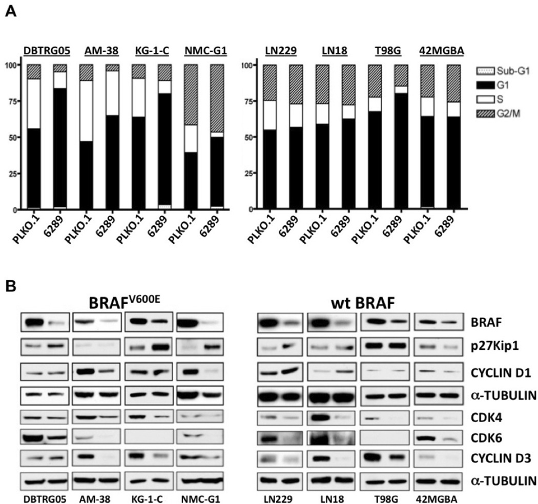
Experimental design: BRAF(V600E) mutation status was examined in two pediatric MA patient cohorts. For functional studies, BRAF(V600E) MA cell lines were used to investigate the effects of BRAF shRNA knockdown in vitro, and to investigate BRAF pharmacologic inhibition in vitro and in vivo.

Results: BRAF(V600E) mutations were identified in 11 and 10% of MAs from two distinct series of tumors (six of 58 cases total). BRAF was expressed in all MA cell lines examined, among which BRAF(V600E) was identified in four instances. Using the BRAF(V600E)-specific inhibitor PLX4720, pharmacologic blockade of BRAF revealed preferential antiproliferative activity against BRAF(V600E) mutant cells in vitro, in contrast to the use of shRNA-mediated knockdown of BRAF, which inhibited cell growth of glioma cell lines regardless of BRAF mutation status. Using orthotopic MA xenografts, we show that PLX4720 treatment decreases tumor growth and increases overall survival in mice-bearing BRAF(V600E) mutant xenografts, while being ineffective, and possibly tumor promoting, against xenografts with wild-type BRAF.

Conclusions: Our results indicate a 10% incidence of activating BRAF(V600E) among pediatric MAs. With regard to implications for therapy, our results support evaluation of BRAF(V600E)-specific inhibitors for treating BRAF(V600E) MA patients.


Conflict of interest statement

We declare no conflict of interest (or relationship that would be suspected of constituting conflicts) at the time of submission.

Figures

Figure 1
Figure 1
BRAF, CRAF, and downstream signaling mediator activation in MA cell lines. A. Cell lysates from 20 human MA cell lines were examined by Western Blot using antibodies against the indicated proteins. Cell lines harboring mutant BRAF are indicated by the dotted line. BRAF protein signals were normalized against corresponding α-TUBULIN signals, with ratios expressed in relation to a normal human astrocyte (Clonetics) value of 100. A second NHA cell source (AllCells) was determined as expressing nearly identical BRAF as the Clonetics NHAs that were used for establishing MA cell line BRAF expression levels.
Figure 2
Figure 2
shRNA suppression of BRAF expression inhibits ERK phosphorylation and in vitro growth of MA cell lines. A. DBTRG-05MG cells were infected with lentivirus expressing empty vector (PLKO.1), or with each of 4 BRAF shRNA lentivirus. Results show that lentivirus 6289 is the most effective at suppressing BRAF expression, as well as being the most effective in suppressing phosphorylation of ERK. B. In vitro growth analysis of modified DBTRG-05MG cells (left) shows decreased growth rate of cells transduced with 6289 lentivirus. Control and 6289 shRNA derivatives for wild-type BRAF cell line 42MGBA were similarly examined for effects of BRAF knockdown on p-ERK (A, right panel) and cell proliferation (B, right panel).
Fig 3
Fig 3
Effect of shRNA mediated BRAF suppression on MA cell cycle distributions and expression of cell cycle regulatory proteins. A. MA cell lines with (left) or without (right) BRAFV600E mutation were infected with lentivirus expressing empty control (PLKO.1) or BRAF shRNA (6289), then treated with puromycin to select infected cells. Pooled puromycin resistant cells were subjected to flow cytometry to determine cell cycle distributions. For all cell lines with BRAFV600E, suppression of BRAF expression resulted in increased proportion of G1 phase cells. For 3 of th 4 cell lines with wild-type BRAF, shRNA suppression had little effect on the G1 fraction of cells. B. Lysates from the same transduced cells as in “A” were subjected to Western Blot analysis to assess the effects of BRAF knockdown on the expression of proteins with known roles in regulating G1 phase cell cycle transit: CYCLIN D1 and CYCLIN D3, enzymatic subunits CDK4 and CDK6, and cyclin dependent kinase inhibitor p27Kip1. The left lane in each blot results from control shRNA and the right lane result is from use of the BRAF shRNA lentivirus. Comparison of results for any two cell lines shows that BRAF suppression effects are variable for the five G1 regulatory proteins examined, but are consistent in demonstrating that BRAF suppression results in decreased expression of one or more of the proteins that promote G1 transit, and/or increases the expression of the negative G1 transit regulator p27Kip1.
Fig 4
Fig 4
Effects of BRAF pharmacologic inhibition on cell proliferation, and on EGF stimulated signal transduction. A. DBTRG-05MG and AM-38, which harbor BRAFV600E, and 42MGBA, with wild-type BRAF, were incubated with increasing concentrations of PLX4720, with effects on growth inhibition indicated after 3 days treatment. EC50 values for DBTRG-05MG and AM-38 are 1.75 and 6.19 µM, respectively, whereas the EC50 for 42MGBA is much higher: 31.20 µM. B. Cultures of DBTRG-05MG, AM-38, and 42MGBA were incubated with serum free medium for 4 hours, then treated with the indicated concentrations of PLX4720 for 1 hour, followed by the addition of 10 ng/ml EGF. Ten min after the addition of EGF, cell lysates were harvested and analyzed by Western blot analysis using the indicated antibodies. Note that a negative effect on p-MEK and p-ERK is evident at 2 and 10 uM PLX4720 for BRAFV600E cells, but not for wild-type BRAF 42MGBA cells. C. GBM cells cultured in the presence of DMSO (0.1%), or 2 or 10 uM PLX4720, for 20 hours, and then harvested for cell cycle analysis with flow cytometry.
Figure 5
Figure 5
Effect of PLX4720 on tumor growth and survival for mice with intracranial MA xenografts. A) Mice received intracranial injection of 300,000 luciferase modified BRAFV600E AM-38 cells, with intraperitoneal administration of vehicle (DMSO) or PLX4720 (20 mg/kg) daily for 2 weeks, beginning at day 7. Survival plot (left) shows that PLX4720 treatment significantly extends the survival of mice with intracranial BRAFV600E tumors, consistent with results from quantitative BLI which show significant anti-proliferative effect of PLX4720 at the first imaging time point subsequent to initiation of treatment (p = 0.018 for control vs. PLX4720 BLI values: middle panel). Bioluminescence images (right) of intracranial tumor in control and PLX4720 treatment group mice show lesser AM-38 bioluminescence signal in the mouse administered PLX4720 following the 5th day of PLX4720 treatment (mice shown had median treatment group luminescence values at time of initiation of therapy). B) In contrast, no PLX4720 survival advantage nor anti-proliferative effect is evident for mice with wild-type BRAF intracranial xenografts (U87).

References

    1. Stupp R, Mason WP, van den Bent MJ, Weller M, Fisher B, Taphoorn MJ, et al. Radiotherapy plus concomitant and adjuvant temozolomide for glioblastoma. N Engl J Med. 2005;352:987–996. - PubMed
    1. Cohen KJ, Heideman RL, Zhou T, Holmes EJ, Lavey RS, Bouffet E, Pollack IF. Temozolomide in the treatment of children with newly diagnosed diffuse intrinsic pontine gliomas: a report from the Children's Oncology Group. Neuro Oncol. 2011;13:410–416. - PMC - PubMed
    1. Cancer Genome Atlas Research Network. Comprehensive genomic characterization defines human glioblastoma genes and core pathways. Nature. 2008;455:1061–1068. - PMC - PubMed
    1. Parsons DW, Jones S, Zhang X, Lin JC, Leary RJ, Angenendt P, et al. An integrated genomic analysis of human glioblastoma multiforme. Science. 2008;321:1807–1812. - PMC - PubMed
    1. Pollack IF, Hamilton RL, James CD, Finkelstein SD, Burnham J, Yates AJ, et al. Rarity of PTEN deletions and EGFR amplification in malignant gliomas of childhood: Results from the children's cancer group 945 cohort. J Neurosurg. 2006;105:418–424. - PubMed

Publication types

MeSH terms