. 2012 Feb;40(4):1828-40.
doi: 10.1093/nar/gkr867. Epub 2011 Oct 27.

Crystal structure of a heterodimer of editosome interaction proteins in complex with two copies of a cross-reacting nanobody

Affiliations

Crystal structure of a heterodimer of editosome interaction proteins in complex with two copies of a cross-reacting nanobody

Young-Jun Park et al. Nucleic Acids Res. 2012 Feb.

Abstract

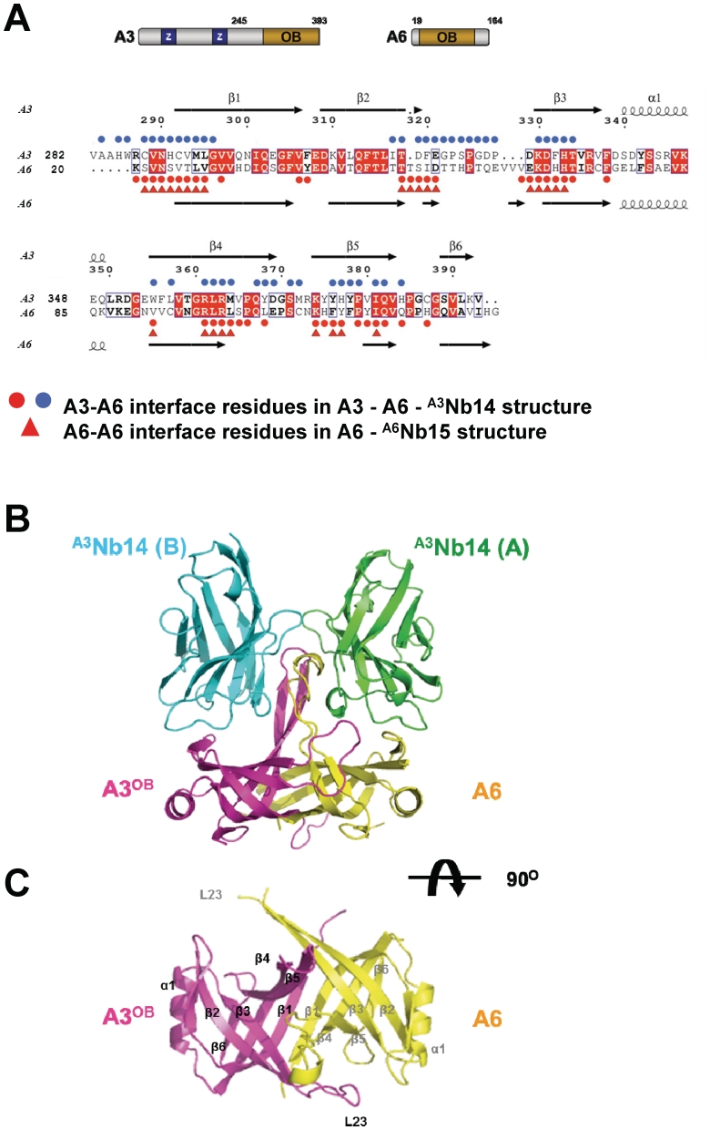
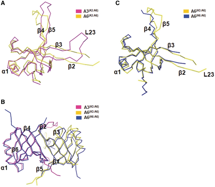
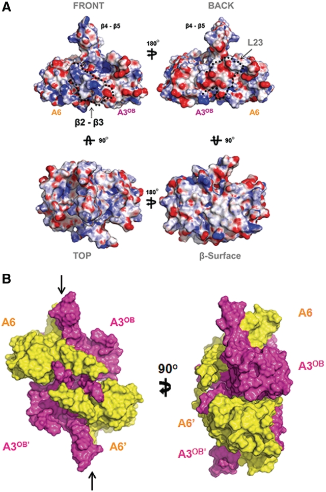
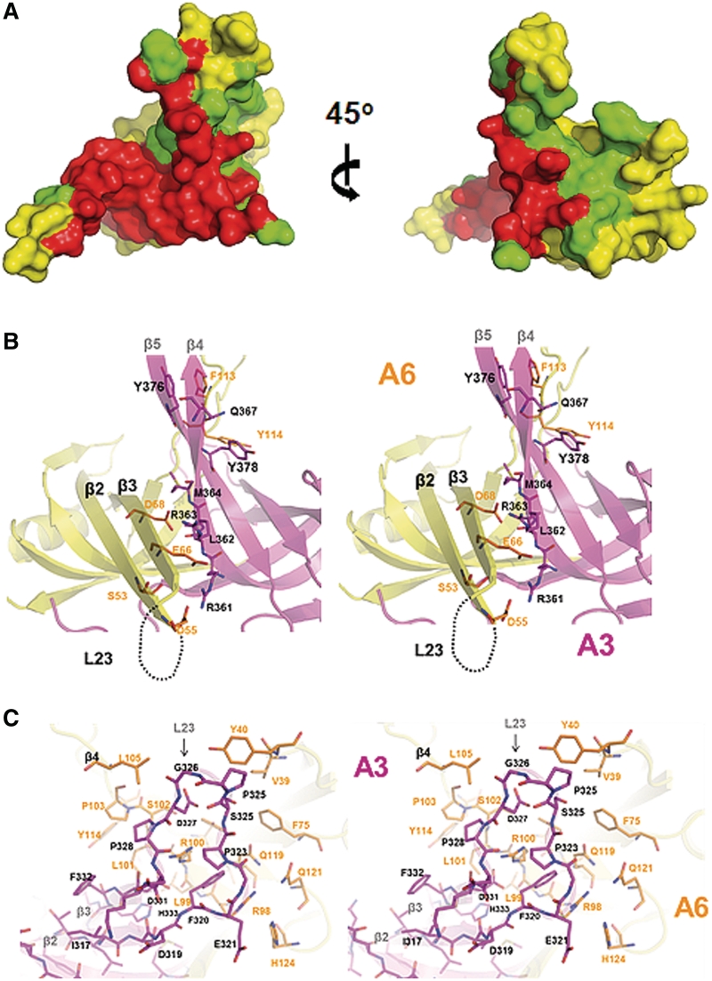
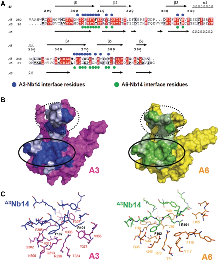
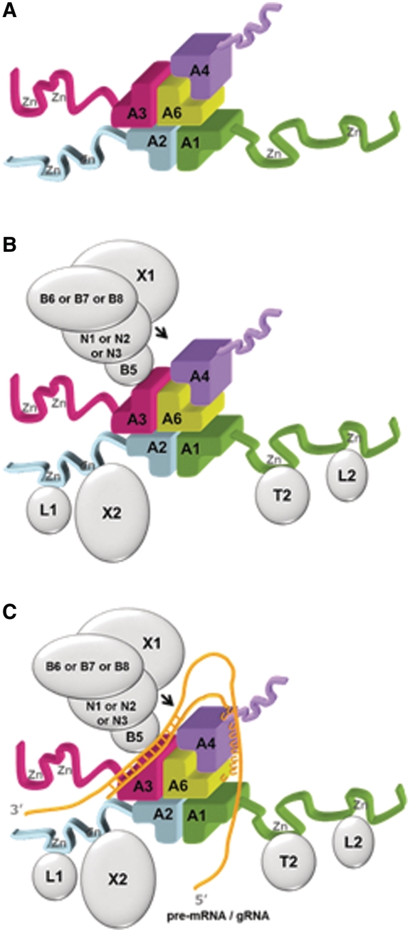
The parasite Trypanosoma brucei, the causative agent of sleeping sickness across sub-Saharan Africa, depends on a remarkable U-insertion/deletion RNA editing process in its mitochondrion. A approximately 20 S multi-protein complex, called the editosome, is an essential machinery for editing pre-mRNA molecules encoding the majority of mitochondrial proteins. Editosomes contain a common core of twelve proteins where six OB-fold interaction proteins, called A1-A6, play a crucial role. Here, we report the structure of two single-strand nucleic acid-binding OB-folds from interaction proteins A3 and A6 that surprisingly, form a heterodimer. Crystal growth required the assistance of an anti-A3 nanobody as a crystallization chaperone. Unexpectedly, this anti-A3 nanobody binds to both A3(OB) and A6, despite only ~40% amino acid sequence identity between the OB-folds of A3 and A6. The A3(OB)-A6 heterodimer buries 35% more surface area than the A6 homodimer. This is attributed mainly to the presence of a conserved Pro-rich loop in A3(OB). The implications of the A3(OB)-A6 heterodimer, and of a dimer of heterodimers observed in the crystals, for the architecture of the editosome are profound, resulting in a proposal of a 'five OB-fold center' in the core of the editosome.


Figures

Figure 1.
Figure 1.
Structure of heterotetramer and heterodimer. (A) Sequence alignment of A3OB and A6. A schematic representation of full-length A3 and full-length A6 with their OB-fold domain and the zinc finger domains (Z) shown in the upper panel. A3OB and A6 sequences (acronyms provided on the left) are aligned in the lower panel. Strictly conserved residues in both sequences are in red boxes. Secondary structure elements are indicated on top and below. Red and blue spheres indicate residues forming the A3OB–A6 interface; red triangles the interface residues of the A6 dimer (PDB-ID: 3K7U) (54). (B) The A3OB–A6-(A3Nb14)2 heterotetramer. The heterotetramer is shown perpendicular to the pseudo-2-fold axis relating the OB-folds of A3OB and A6. A3OB is depicted in magenta; A6 in yellow; A3Nb14 in complex with A3OB in blue; A3Nb14 in complex with A6 in green. The heterotetramer depicted is part of an [A3OB–A6–(A3Nb14)2]2 heterooctamer occurring in the crystal lattice (Supplementary Figure S7). (C) The A3OB–A6-heterodimer. A view along the pseudo-2-fold of the A3OB–A6 dimer of the heterotetramer shown in (B) is in the same color code. The N-terminal β-strands β1 of each monomer run anti-parallel to each other in the center of an extended β-sheet.
Figure 2.
Figure 2.
Comparison of A3OB and A6. (A) Superposition of the A3OB and A6 chains from the A3OB-A6 heterodimer. The A3OB chain is shown in magenta; A6 from the A3OB–A6 heterodimer in yellow. Note the large conformational differences in the L23 loop, as well as in the β4–β5 hairpin. (B) Superposition of the A3OB–A6 and A6–A6 dimers. A view along the pseudodyad of the heterodimer. The A3OB chain is shown in magenta; A6 from the A3OB–A6 heterodimer in yellow; both A6 subunits from the A6–A6 homodimer in blue (PDB-ID: 3K7U) (54). (C) Superposition of A6 in the A3OB-A6 heterodimer and A6 in the A6 homodimer. A6 from the heterodimer is depicted in yellow; A6 from the homodimer in blue (PDB-ID: 3K7U) (54). Note that the L23 loop, as well as the β4–β5 hairpin of A6 is better ordered in the A3OB-A6 heterodimer than in the A6 dimer.
Figure 3.
Figure 3.
Characteristics of A3OB–A6 heterodimer and heterotetramer. (A) Surface charge distribution. Four views of the surface charge distribution of the A3OB–A6 heterodimer. The electrostatic potential surface of the A3OB and A6 dimer is calculated using APBS (71) and displayed as blue for a positively charged surface potential, red for negative and gray for neutral. The ‘front surface’ (upper left) is predominantly positively charged and the ‘back surface’ (upper right) predominantly negative. The ‘top’ view (lower left) shows pockets of positively charged surface areas next to the protruding β4–β5 hairpin. The ‘β-surface’ (lower right), formed mainly by the extended β-sheet of the dimer, contains large hydrophobic areas (gray). (B) The ‘shifted tetramer’ formed by two A3OB-A6 heterodimers in the crystal. The A3OB and A6 proteins in the complex are shown in surface mode, with the two A3OB chains in magenta and the two A6 domains in yellow. The β-surfaces of two A3OB–A6 heterodimers contact each other in the crystals. The ∼2000 Å2 total solvent accessible surface buried in this dimer–dimer interface is composed of ∼800 Å2 from A3OB–A3OB′ contacts, ∼600 Å2 contributed by A3OB–A6, ∼600 Å2 by A3OB′–A6. The A6 and A6 subunits do not interact, while A3OB interacts with three different OB-folds in this assembly. This (A3OB–A6)2 heterotetramer is distinct from canonical OB-fold homotetramers (78) in that the upper and lower dimer pseudo-dyads are not coinciding, but are shifted with respect to each other parallel to the interacting β-surface (See arrows representing pseudo-dyads in the upper and lower heterodimers).
Figure 4.
Figure 4.
Interactions in A3OB–A6 compared with those in A6–A6. (A) The fingerprint of A3OB on A6 is larger than that of A6 on A6. The interactions are ‘painted’ onto the surface of A6. Red: the contact area which is the same in the A3OB–A6 heterodimer and in the A6 homodimer. Green: the extra contact area in the heterodimer compared with the homodimer. Yellow: the surface of A6 neither in contact with A3OB in the heterodimer, nor with the second A6 subunit in the homodimer. The surface buried in the interaction in A3OB–A6 is ∼35% larger than in the A6–A6 dimer (PDB-ID: 3K7U)(54). (B) Strands β2–β3 of A6 contacting A3OB. A stereo view of the ‘second contact area’ (see text) of A3–A6 with important residues highlighted shown in ribbon representation with the same color coding as in Figure 1B. The secondary structure elements and key residues mediating the A3–A6 dimer interactions are labeled. (C) The Pro-rich L23 loop of A3OB contacting A6. A stereo view, 180° different about the vertical axis from (B), of the ‘third contact area’ (see text) in the heterodimer shown with stick representations. Selected secondary structure elements and key contact residues are labeled. A3 is colored magenta, A6 yellow-gold.
Figure 5.
Figure 5.
The A3OBA3Nb14 and A6–A3Nb14 complexes. (A) Sequences and interface residues of A3OB and A6 with A3Nb14. Structure-based alignment of the T. brucei A3 sequence (upper line) and A6 sequence (lower line). Residues highlighted in red are completely conserved. Blue spheres indicate A3 residues in contact with nanobody A3Nb14. Green sphere residues of A6 are residues in contact with the second copy of nanobody A3Nb14 in the heterotetramer. (B) Footprints of A3Nb14 onto A3OB (left) and onto A6 (right). Interaction Region 1 is outlined by an ellipse with a solid line; Interaction Region 2 with a dashed line. Left: dark blue: A3OB residues contacting A3Nb14 residues which are identical in A6; Light blue: A3OB residues contacting A3Nb14 residues which are different in A6; Magenta: A3OB atoms not contacting the nanobody. Right: dark green: A6 residues contacting A3Nb14 residues which are identical in A3OB; light green: A3Nb14 residues-contacting residues of A6 which are different in A3OB. Yellow: A6 atoms not contacting the nanobody. (C) Key interactions of A3Nb14 with A3 (left) and of A3Nb14 with A6 (right). Close-ups of Interaction Region 1 of both A3 and A6 are shown in stick representations. Residues involved in protein–protein interactions are indicated and colored according to the molecules they belong to. Colors: A3OB in magenta with its A3Nb14 bound in blue, A6 in yellow-gold with its A3Nb14 bound in green. Nitrogens blue, oxygens red.
Figure 6.
Figure 6.
An OB-fold center in the core of the editosome. (A) Model for a “five OB-fold” center of the editosome. The basis is a shifted heterotetramer (in this example, comprising the OB-folds of A1, A2, A3 and A6) plus an additional A4OB fold interacting with A6. Please note that while the A3OB–A6 heterodimer is experimentally observed, the positions of A1OB, A2OB and A4OB in this figure can be interchanged and still yield a model in agreement with the experimental constraints mentioned in the text. Therefore, the arrangement shown is one of six possible models of these five OB-folds forming the center of the editosome core. (B) The same five OB-fold center as in (A) with the additional domains in the OB-fold interaction proteins shown as ribbons and the proteins interacting with these domains outlined as silver ellipsoids. Note that three different types of editosomes have been reported to exist, containing either N1-B6, or N2-B7, or N3-B8-X1, in addition to the core (52). (C) A global outline of how a pre-mRNA•gRNA duplex might interact with the five OB-fold center shown in (A). The gold wires indicate RNA, with the duplex in the anchor region shown as crossbars, and the poly-U tail of the mature gRNA as a series of U's. The arrow points to an editing site. The surrounding enzymes might display mobility with respect to the five OB-fold center. See also text.

References

    1. Croft SL, Sundar S, Fairlamb AH. Drug resistance in leishmaniasis. Clin. Microbiol. Rev. 2006;19:111–126. - PMC - PubMed
    1. Fairlamb AH. Chemotherapy of human African trypanosomiasis: current and future prospects. Trends. Parasitol. 2003;19:488–494. - PubMed
    1. Hotez PJ, Molyneux DH, Fenwick A, Kumaresan J, Sachs SE, Sachs JD, Savioli L. Control of neglected tropical diseases. N. Engl. J. Med. 2007;357:1018–1027. - PubMed
    1. Tarleton RL, Reithinger R, Urbina JA, Kitron U, Gurtler RE. The challenges of Chagas Disease–grim outlook or glimmer of hope. PLoS Med. 2007;4:e332. - PMC - PubMed
    1. Moyersoen J, Choe J, Fan E, Hol WG, Michels PA. Biogenesis of peroxisomes and glycosomes: trypanosomatid glycosome assembly is a promising new drug target. FEMS Microbiol. Rev. 2004;28:603–643. - PubMed

Publication types

Associated data