. 2011;6(10):e26047.
doi: 10.1371/journal.pone.0026047. Epub 2011 Oct 19.

FilmArray, an automated nested multiplex PCR system for multi-pathogen detection: development and application to respiratory tract infection

Affiliations

FilmArray, an automated nested multiplex PCR system for multi-pathogen detection: development and application to respiratory tract infection

Mark A Poritz et al. PLoS One. 2011.

Erratum in

  • PLoS One. 2011;6(11). doi: 10.1371/annotation/468cfdcd-184c-42f7-a1d0-3b72a2f6a558

Abstract

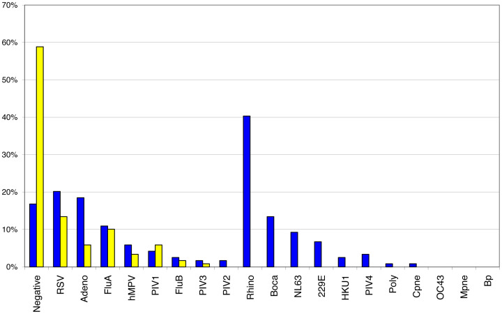
The ideal clinical diagnostic system should deliver rapid, sensitive, specific and reproducible results while minimizing the requirements for specialized laboratory facilities and skilled technicians. We describe an integrated diagnostic platform, the "FilmArray", which fully automates the detection and identification of multiple organisms from a single sample in about one hour. An unprocessed biologic/clinical sample is subjected to nucleic acid purification, reverse transcription, a high-order nested multiplex polymerase chain reaction and amplicon melt curve analysis. Biochemical reactions are enclosed in a disposable pouch, minimizing the PCR contamination risk. FilmArray has the potential to detect greater than 100 different nucleic acid targets at one time. These features make the system well-suited for molecular detection of infectious agents. Validation of the FilmArray technology was achieved through development of a panel of assays capable of identifying 21 common viral and bacterial respiratory pathogens. Initial testing of the system using both cultured organisms and clinical nasal aspirates obtained from children demonstrated an analytical and clinical sensitivity and specificity comparable to existing diagnostic platforms. We demonstrate that automated identification of pathogens from their corresponding target amplicon(s) can be accomplished by analysis of the DNA melting curve of the amplicon.


Conflict of interest statement

Competing Interests: I have read the journal's policy and have the following conflicts: Several authors are current or former employees of Idaho Technology. PCT Applications WO2006/121997 and WO2008/140568 have been filed on aspects of the work described here. AJB, JD, AH and CLB collaborate with Idaho Technology, Inc., on several National Institutes of Health and Centers for Disease Control-funded projects (see funding). There are no other relevant declarations relating to employment, consultancy, patents, products in development or modified products. This does not alter our adherence to all the PLoS ONE policies on sharing data and materials.

Figures

Figure 1
Figure 1. FilmArray pouch.
(A) A FilmArray pouch was injected with mock sample (here colored blue for illustrative purposes) in the left side injection port and hydration solution (colored red) in the right side injection port. (B) The blisters of a FilmArray pouch were filled with different coloring (and the channels between the blisters heat sealed shut). In this pouch the plunger tree was made from plastic dyed blue. The fitment and film are normally at right angles to each other; for clarity the pouch has been flattened. (C) A schematic of the pouch showing a trace of the blisters, channels and array wells (black) and the functional areas of the pouch (red).
Figure 2
Figure 2. Schematic of second stage PCR mix entering the array.
The layers of film and adhesive attaching the array to the pouch are separated to show the flow of liquid into the cells of the array (figure is not to scale). From the top the layers are: 2nd pouch film, 1st pouch film, array adhesive layer (orange), pricked cover film, array (black, with wells), and array cover film. All of the actual layers are transparent except for the array itself. Second stage PCR primers are spotted into the cells during manufacture and air-dried (Methods). Arrows show the flow of PCR master mix (without primers) entering the array through a hole cut in the 1st pouch film.
Figure 3
Figure 3. FilmArray instrument with pouch being loaded.
Figure 4
Figure 4. Real-time amplification and melt curves from the array.
Respiratory Pathogen pouches were injected with viral transport medium spiked with 200 TCID50 FluA H1-seasonal (panels A and B), 4×10⁶ cfu B. per and 200 TCID50 FluA-H1 (panels C and D), or 4×10⁶ cfu B. per (panels E and F) and run on the FilmArray instrument. Real time amplification curves (panels A and C and E) and post-amplification melt curves (panels B and D and F) for selected wells on the array are shown. Assays are spotted in triplicate: FluA-pan1 (orange), FluA-pan2 (pink), FluA-H1-pan (red), FluA-H3 (black), B. per (Green), Yeast RNA process control (dark blue), Second stage PCR control (light blue). For clarity the controls are shown in panels E and F only.
Figure 5
Figure 5. Detection rates of the FilmArray RP pouch compared to DFA.
Pediatric NPA samples (N = 328) were tested either by DFA at PCMC (yellow bars) or on the FilmArray (Blue bars). The percent of samples in which no virus (Negative) or one of the indicated viruses was detected is shown. The viruses are grouped into those in which both DFA and FilmArray assays are available or only the FilmArray assay is available.
Figure 6
Figure 6. Amplification and melt curves at low target levels.
Respiratory Pathogen pouches were injected with viral transport medium spiked with 1 TCID50 of the FluA- H1 seasonal virus used in Figure 4, and run on the FilmArray instrument. Real-time amplification curves (A) and post-amplification melt curves (B) for selected wells on the array are shown. Assays are spotted in triplicate: FluA-pan1 (orange), FluA-pan2 (pink), FluA-H1-pan (red). The ordinate scales are the same as in Figure 4.
Figure 7
Figure 7. Tm data used to establish assay specific melt windows.
Histograms of the theoretical or observed Tms of the hMPV assay are shown. Tm data for the FilmArray runs includes each of the three replicates of the second stage PCR. A: Tms calculated from 13 sequence variants published in the NCBI databases. B: Tm data generated during the system beta-testing with 37 banked hMPV-positive patient samples. C: Tm data generated during the inclusivity testing with 10 hMPV strains representing subtypes A1, A2, B1 and B2. Multiple FilmArray runs of these strains are included in this data set. D: Tm data from 74 hMPV-positive patient samples collected during the clinical evaluation.

References

    1. Zhang ZW, Zhou YM, Zhang Y, Guo Y, Tao SC, et al. Sensitive detection of SARS coronavirus RNA by a novel asymmetric multiplex nested RT-PCR amplification coupled with oligonucleotide microarray hybridization. Methods Mol Med. 2005;114:59–78. - PMC - PubMed
    1. Petric M, Comanor L, Petti CA. Role of the laboratory in diagnosis of influenza during seasonal epidemics and potential pandemics. J Infect Dis. 2006;194(Suppl 2):S98–110. - PubMed
    1. Fox JD. Respiratory virus surveillance and outbreak investigation. J Clin Virol. 2007;40(Suppl 1):S24–30. - PMC - PubMed
    1. Lessa FC, Gould PL, Pascoe N, Erdman DD, Lu X, et al. Health care transmission of a newly emergent adenovirus serotype in health care personnel at a military hospital in Texas, 2007. J Infect Dis. 2009;200:1759–1765. - PubMed
    1. Dundas NE, Ziadie MS, Revell PA, Brock E, Mitui M, et al. A lean laboratory: operational simplicity and cost effectiveness of the Luminex xTAG respiratory viral panel. J Mol Diagn. 2011;13:175–179. - PMC - PubMed

Publication types

MeSH terms