

Presence of cartilage stem/progenitor cells in adult mice auricular perichondrium
- PMID: 22039478
- PMCID: PMC3198405
- DOI: 10.1371/journal.pone.0026393
Presence of cartilage stem/progenitor cells in adult mice auricular perichondrium
Abstract
Background: Based on evidence from several other tissues, cartilage stem/progenitor cells in the auricular cartilage presumably contribute to tissue development or homeostasis of the auricle. However, no definitive studies have identified or characterized a stem/progenitor population in mice auricle.
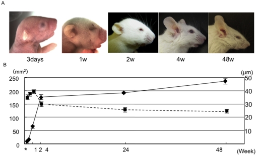
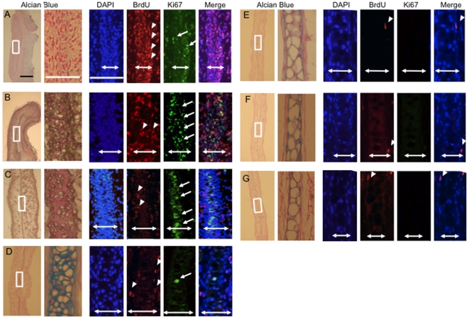
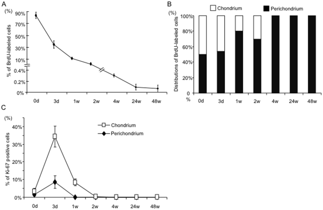
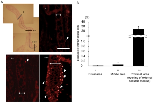
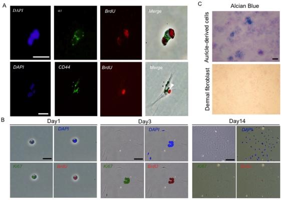
Methodology/principal findings: The 5-bromo-2'-deoxyuridine (BrdU) label-retaining technique was used to label dividing cells in fetal mice. Observations one year following the labeling revealed that label-retaining cells (LRCs) were present specifically in auricular perichondrium at a rate of 0.08±0.06%, but LRCs were not present in chondrium. Furthermore, LRCs were successfully isolated and cultivated from auricular cartilage. Immunocytochemical analyses showed that LRCs express CD44 and integrin-α(5). These LRCs, putative stem/progenitor cells, possess clonogenicity and chondrogenic capability in vitro.
Conclusions/significance: We have identified a population of putative cartilage stem/progenitor cells in the auricular perichondrium of mice. Further characterization and utilization of the cell population should improve our understanding of basic cartilage biology and lead to advances in cartilage tissue engineering and novel therapeutic strategies for patients with craniofacial defects, including long-term tissue restoration.
Conflict of interest statement
Figures
Similar articles
-
Reconstruction of human elastic cartilage by a CD44+ CD90+ stem cell in the ear perichondrium.Proc Natl Acad Sci U S A. 2011 Aug 30;108(35):14479-84. doi: 10.1073/pnas.1109767108. Epub 2011 Aug 11. Proc Natl Acad Sci U S A. 2011. PMID: 21836053 Free PMC article.
-
Identification of cartilage progenitor cells in the adult ear perichondrium: utilization for cartilage reconstruction.Lab Invest. 2006 May;86(5):445-57. doi: 10.1038/labinvest.3700409. Lab Invest. 2006. PMID: 16625212
-
The in vivo chondrogenesis of cartilage stem/progenitor cells from auricular cartilage and the perichondrium.Am J Transl Res. 2019 May 15;11(5):2855-2865. eCollection 2019. Am J Transl Res. 2019. PMID: 31217859 Free PMC article.
-
Regenerative Potential of Perichondrium: A Tissue Engineering Perspective.Tissue Eng Part B Rev. 2022 Jun;28(3):531-541. doi: 10.1089/ten.TEB.2021.0054. Epub 2021 Jun 30. Tissue Eng Part B Rev. 2022. PMID: 33966486 Review.
-
[Regenerative medicine of tissue engineering: auricular cartilage regeneration and functional reconstruction].Lin Chuang Er Bi Yan Hou Tou Jing Wai Ke Za Zhi. 2019 Jun;33(6):567-571. doi: 10.13201/j.issn.1001-1781.2019.06.024. Lin Chuang Er Bi Yan Hou Tou Jing Wai Ke Za Zhi. 2019. PMID: 31163539 Review. Chinese.
Cited by
-
Meis2 controls skeletal formation in the hyoid region.Front Cell Dev Biol. 2022 Sep 28;10:951063. doi: 10.3389/fcell.2022.951063. eCollection 2022. Front Cell Dev Biol. 2022. PMID: 36247013 Free PMC article.
-
PPARD is an Inhibitor of Cartilage Growth in External Ears.Int J Biol Sci. 2017 May 16;13(5):669-681. doi: 10.7150/ijbs.19714. eCollection 2017. Int J Biol Sci. 2017. PMID: 28539839 Free PMC article.
-
Oriented clonal cell dynamics enables accurate growth and shaping of vertebrate cartilage.Elife. 2017 Apr 17;6:e25902. doi: 10.7554/eLife.25902. Elife. 2017. PMID: 28414273 Free PMC article.
-
Modeling Normal and Pathological Ear Cartilage in vitro Using Somatic Stem Cells in Three-Dimensional Culture.Front Cell Dev Biol. 2020 Jul 28;8:666. doi: 10.3389/fcell.2020.00666. eCollection 2020. Front Cell Dev Biol. 2020. PMID: 32850801 Free PMC article.
-
Head to Knee: Cranial Neural Crest-Derived Cells as Promising Candidates for Human Cartilage Repair.Stem Cells Int. 2019 Jan 15;2019:9310318. doi: 10.1155/2019/9310318. eCollection 2019. Stem Cells Int. 2019. PMID: 30766608 Free PMC article. Review.
References
-
- Bickenbach JR, Chism E. Selection and extended growth of murine epidermal stem cells in culture. Exp Cell Res. 1998;244:184–195. - PubMed
-
- Brandl C, Florian C, Driemel O, Weber BH, Morsczeck C. Identification of neural crest-derived stem cell-like cells from the corneal limbus of juvenile mice. Exp Eye Res. 2009;89:209–217. - PubMed
-
- Zuk PA, Zhu M, Mizuno H, Huang J, Futrell JW, et al. Multilineage cells from human adipose tissue: implications for cell-based therapies. Tissue Eng. 2001;7:211–228. - PubMed
-
- Bairati A, Comazzi M, Gioria M. A comparative study of perichondrial tissue in mammalian cartilages. Tissue Cell. 1996;28:455–468. - PubMed
Publication types
MeSH terms
Substances
LinkOut - more resources
Full Text Sources
Other Literature Sources
Medical
Miscellaneous
