. 2011;6(10):e26432.
doi: 10.1371/journal.pone.0026432. Epub 2011 Oct 19.

Targeted decorin gene therapy delivered with adeno-associated virus effectively retards corneal neovascularization in vivo

Affiliations

Targeted decorin gene therapy delivered with adeno-associated virus effectively retards corneal neovascularization in vivo

Rajiv R Mohan et al. PLoS One. 2011.

Abstract

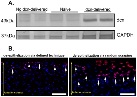
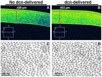
Decorin, small leucine-rich proteoglycan, has been shown to modulate angiogenesis in nonocular tissues. This study tested a hypothesis that tissue-selective targeted decorin gene therapy delivered to the rabbit stroma with adeno-associated virus serotype 5 (AAV5) impedes corneal neovascularization (CNV) in vivo without significant side effects. An established rabbit CNV model was used. Targeted decorin gene therapy in the rabbit stroma was delivered with a single topical AAV5 titer (100 µl; 5×10(12) vg/ml) application onto the stroma for two minutes after removing corneal epithelium. The levels of CNV were examined with stereomicroscopy, H&E staining, lectin, collagen type IV, CD31 immunocytochemistry and CD31 immunoblotting. Real-time PCR quantified mRNA expression of pro- and anti-angiogenic genes. Corneal health in live animals was monitored with clinical, slit-lamp and optical coherence tomography biomicroscopic examinations. Selective decorin delivery into stroma showed significant 52% (p<0.05), 66% (p<0.001), and 63% (p<0.01) reduction at early (day 5), mid (day 10), and late (day 14) stages of CNV in decorin-delivered rabbit corneas compared to control (no decorin delivered) corneas in morphometric analysis. The H&E staining, lectin, collagen type IV, CD31 immunostaining (57-65, p<0.5), and CD31 immunoblotting (62-67%, p<0.05) supported morphometric findings. Quantitative PCR studies demonstrated decorin gene therapy down-regulated expression of VEGF, MCP1 and angiopoietin (pro-angiogenic) and up-regulated PEDF (anti-angiogenic) genes. The clinical, biomicroscopy and transmission electron microscopy studies revealed that AAV5-mediated decorin gene therapy is safe for the cornea. Tissue-targeted AAV5-mediated decorin gene therapy decreases CNV with no major side effects, and could potentially be used for treating patients.


Conflict of interest statement

Competing Interests: The authors have declared that no competing interests exist.

Figures

Figure 1
Figure 1. Representative immunoblot (A) and TUNEL assay (B) of rabbit corneas receiving AAV5-decorin or AAV5-gfp.
Representative western blotting showing AAV5-controlled decorin delivery in rabbit corneas collected 14 days after VEGF-implantation (A) and TUNEL assay detecting keratocyte apoptosis in rabbit corneas collected 4 hours after epithelial removal by two different ways (B). A strong band in AAV5-dcn treated rabbit corneas of day-14 time point reveals significant decorin delivery (8.7-fold, p<0.05) in the stroma, and a weak decorin band in naive or AAV5-gfp treated corneas indicates low endogenous decorin expression (A). GAPDH was used to confirm equal protein loading in each well and normalization of the data. TUNEL-stained rabbit corneal sections of 4-hour time point shown in Panel B demonstrate minimal keratocyte apoptosis (arrow) in cornea deepithelialized via gentle scrapping with #64 surgical blade by running blade at 45° angle (left panel) compared to high keratocyte death in cornea in which epithelium was removed in random manner with #64 surgical blade (right panel). Scale bar denotes 100 µm. dcn = decorin.
Figure 2
Figure 2. Representative stereomicroscopy images showing VEGF-induced CNV in no decorin-delivered control (A, C and E) and decorin-delivered (B, D and F) rabbit corneas.
Rabbit eyes were imaged early (5 day, Panels A, B), mid (10 day, Panels C, D), and late (14 day, Panels E, F) stages after VEGF pellet implantation. The 100 µl AAV5 viral titer (5×10¹² vg/ml) expressing no gene/gfp or decorin was topically applied onto the cornea after removing corneal epithelium for single application for 2 minutes. A statistically significant inhibition of neovascularization was observed at three tested early, mid and late stages of the CNV. Scale bar denotes 2 mm. dcn = decorin.
Figure 3
Figure 3. Morphometric quantification of CNV from different time points in rabbit eyes receiving AAV5+/−decorin.
Data was collected after day 5 (early), 10 (mid), and 14 (late) of VEGF implantation from the control (no decorin-delivered) and decorin-delivered rabbit corneas. AAV-mediated decorin gene therapy demonstrated statistically significant decrease at the three tested stages of the CNV in rabbit model in vivo. ψ = p<0.05, * = p<0.001, ζ = p<0.01 compared to control. dcn = decorin.
Figure 4
Figure 4. Representative H&E staining images showing anti-angiogenic efficiency of decorin gene therapy in rabbit corneas.
The decorin-delivered rabbit corneas (B and D) showed a statistically significant (p<0.01) reduction in vasculature area, density, and blood vessels number, length and diameter compared to control (no decorin-delivered) rabbit corneas (A and C) collected 14 days after VEGF implantation. The images shown in panels A and B are from the corneal sections obtained from the peripheral region of the cornea closer to the limbus whereas the images shown in panels C and D are from the corneal sections proximal to the VEGF pellet. Scale bar denotes 100 µm. dcn = decorin.
Figure 5
Figure 5. Representative images showing CNV in rabbit corneas treated AAV5+/−decorin.
Lectin (A–F) and collagen type IV (G–L) immunostaining was carried out in corneal tissue sections obtained from AAV5-gfp and AAV5-dcn treated rabbit corneas collected 14 days after VEGF implantation. As expected very high lectin (A–C) and collagen type IV (G–I) immunostaining was detected in the corneal sections of AAV5-gfp-treated (no decorin-delivered) corneas due CNV induced by the VEGF implantation. The AAV5-mediated decorin gene therapy significantly reduced (p<0.01) the CNV as evident from the markedly less lectin+ (D–F) and collagen type IV+ (J–L) cells in decorin-delivered rabbit corneal sections. DAPI stained nuclei are shown in blue whereas lectin and collagen type IV+ cells in red. Scale bar denotes 100 µm. dcn = decorin.
Figure 6
Figure 6. CD31 Western blot for naive, AAV5+/−decorin-delivered rabbit corneal tissues collected 10 and 14 days after VEGF-implantation.
Panel on left side shows immunoblot and on right side shows quantification of western blotting data collected from corneal tissues of day-10 and day-14. A significant decrease in expression of CD31 on day-10 (62%, p<0.05) and day-14 (66%, p<0.05) was detected in decorin-delivered corneas compared to control corneas suggesting that AAV5-mediated decorin gene therapy is efficient in decreasing CNV. β-actin was used to confirm equal loading of protein in each well and normalization of the data. dcn = decorin.
Figure 7
Figure 7. Quantification of mRNA expression of pro- and anti-angiogenic genes by real-time PCR.
Real-time PCR was carried out in naive, control (no decorin-delivered) and decorin-delivered rabbit corneal tissues collected 14 days after VEGF implantation. Decorin gene therapy delivered with AAV5 showed marked decrease in the mRNA expression of VEGF and MCP-1 and less pronounced alteration in angiopoietin and PEDF genes. Our data suggests anti-angiogenic effects of decorin gene therapy are mediated by the down-regulation of pro-angiogeneic (VEGF, MCP1 and angiopoietin) and up-regulation of anti-angiogenic (PEDF) genes. * = p<0.001 compared to naive; ζ = p<0.05 compared to naive; ψ = p<0.05 compared to no decorin (dcn)-delivered.
Figure 8
Figure 8. Representative optical coherence tomography (A and B) and transmission electron microscopy (C and D) images of rabbits receiving AAV5-gfp or AAV5-dcn.
Optical coherence tomography was performed in live rabbits on day 10 after the AAV5-gfp or AAV5-dcn application and transmission electron microscopy was performed in corneas collected 14 days after AAV5-gfp or AAV5-dcn application. The optical scans of no decorin-delivered (A) and decorin-delivered (B) cornea suggest that tissue-selective targeted delivery of decorin gene with AAV5 in the rabbit stroma does not induce changes in corneal structure, thickness or shape. The TEM did not detect significant differences in collagen fibril diameter and arrangement in the naive (C) and decorin-delivered rabbit corneas (D) indicating that localized therapeutic levels of decorin delivery in the cornea does not appear compromise corneal health. dcn = decorin.

References

    1. Ellenberg D, Azar DT, Hallak JA, Tobaigy F, Han KY, et al. Novel aspects of corneal angiogenic and lymphangiogenic privilege. Prog Retin Eye Res. 2010;29:208–248. - PMC - PubMed
    1. Qazi Y, Maddula S, Ambati BK. Mediators of ocular angiogenesis. J Genet. 2009;88:495–515. - PMC - PubMed
    1. Lee P, Wang CC, Adamis AP. Ocular neovascularization: an epidemiologic review. Surv Ophthalmol. 1998;43:245–269. - PubMed
    1. Aydin E, Kivilcim M, Peyman GA, Esfahani MR, Kazi AA, et al. Inhibition of experimental angiogenesis of cornea by various doses of doxycycline and combination of triamcinolone acetonide with low-molecular-weight heparin and doxycycline. Cornea. 2008;27:446–453. - PubMed
    1. Dastjerdi MH, Al-Arfaj KM, Nallasamy N, Hamrah P, Jurkunas UV, et al. Topical bevacizumab in the treatment of corneal neovascularization: results of a prospective, open-label, noncomparative study. Arch Ophthalmol. 2009;127:381–389. - PMC - PubMed

Publication types