. 2011;6(10):e26609.
doi: 10.1371/journal.pone.0026609. Epub 2011 Oct 24.

Tau enhances α-synuclein aggregation and toxicity in cellular models of synucleinopathy

Affiliations

Tau enhances α-synuclein aggregation and toxicity in cellular models of synucleinopathy

Nahuai Badiola et al. PLoS One. 2011.

Abstract

Background: The simultaneous accumulation of different misfolded proteins in the central nervous system is a common feature in many neurodegenerative diseases. In most cases, co-occurrence of abnormal deposited proteins is observed in different brain regions and cell populations, but, in some instances, the proteins can be found in the same cellular aggregates. Co-occurrence of tau and α-synuclein (α-syn) aggregates has been described in neurodegenerative disorders with primary deposition of α-syn, such as Parkinson's disease and dementia with Lewy bodies. Although it is known that tau and α-syn have pathological synergistic effects on their mutual fibrillization, the underlying biological effects remain unclear.

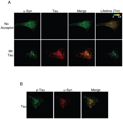
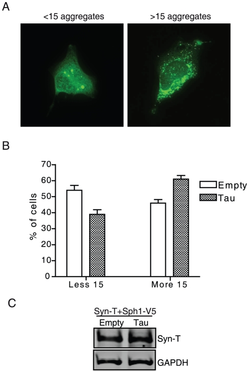
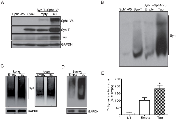
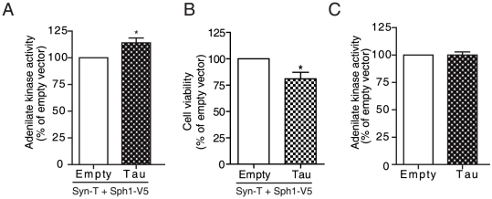
Methodology/principal findings: We used different cell models of synucleinopathy to investigate the effects of tau on α-syn aggregation. Using confocal microscopy and FRET-based techniques we observed that tau colocalized and interacted with α-syn aggregates. We also found that tau overexpression changed the pattern of α-syn aggregation, reducing the size and increasing the number of aggregates. This shift was accompanied by an increase in the levels of insoluble α-syn. Furthermore, co-transfection of tau increased secreted α-syn and cytotoxicity.

Conclusions/significance: Our data suggest that tau enhances α-syn aggregation and toxicity and disrupts α-syn inclusion formation. This pathological synergistic effect between tau and α-syn may amplify the deleterious process and spread the damage in neurodegenerative diseases that show co-occurrence of both pathologies.


Conflict of interest statement

Competing Interests: The authors have declared that no competing interests exist.

Figures

Figure 1
Figure 1. Tau and phospho-tau colocalize and interact with α-syn aggregates in H4 neuroglioma cells.
H4 cells were co-transfected with syn-T plus synphilin1-V5 and empty vector or tau. A, After 48 h cells were fixed and immunostained for α-syn and tau with Alexa Fluor 488 and Alexa 555-labeled antibodies, respectively. Images were analyzed by confocal microscopy and FLIM. Tau and α-syn colocalize in some aggregates but not in all of them (bottom panels). Fluorescence lifetime values are shown in the pseudocolored image. The closeness between α-syn and tau in some aggregates is reflected by a shorter lifetime of Alexa Fluor 488 in the presence of tau. This is represented as a shift from red-yellow towards the green-blue pseudocolor. B, H4 cells were fixed and immunostained for phospho-tau and α-syn with Alexa Fluor 488 and Alexa 555-conjugated antibodies, respectively. Colocalization between phospho-tau and α-syn was observed in some aggregates.
Figure 2
Figure 2. Tau colocalizes and interacts with α-syn aggregates in mouse cortical neurons.
Cortical neurons were co-transfected after 7 days in vitro with α-syn-GFP and tau-RFP. Neurons were then fixed and immunostained for α-syn and tau with Alexa Fluor 488 and Alexa Fluor 555-conjugated antibodies, respectively. Images were analyzed by confocal microscopy and FLIM. The close proximity between α-syn and tau is reflected by a shift towards the green-blue color. The shortening in fluorescence lifetime is particularly evident in some aggregates (arrows).
Figure 3
Figure 3. Overexpression of tau shifts the pattern of α-syn aggregation.
H4 cells were co-transfected with syn-T plus synphilin1-V5 and empty vector or tau. Co-transfection with tau in this model led to an increase in the number of inclusions and a reduction in their size. A, Cells were classified in two groups: cells with less than 15 aggregates and cells with more than 15 aggregates. B, Co-transfection of cells with tau increased the percentage of cells with >15 aggregates as compared with those transfected with empty vector (n = 250 cells). Results are mean ± SEM of 4 independent experiments. C, Total levels of syn-T were measured by western blot. GAPDH was used as a loading control. A representative experiment is shown (n = 4 independent experiments).
Figure 4
Figure 4. Tau increases insoluble α-syn, high molecular weight species and extracellular α-syn levels.
H4 cells were co-transfected with syn-T plus syphilin1-V5 and empty vector or tau. After 48 h cells were lysed and the insoluble fraction was resolved in SDS-PAGE (A) or native-PAGE (B) gels. When tau was co-transfected, we observed an increase in the levels of insoluble α-syn and HMW species of α-syn. GAPDH was used as a loading control. A representative experiment is shown (n = 4 independent experiments). C, Stable H4 cells expressing α-syn constructs (GN-link-aSyn and aSyn-GC) were transfected with either empty vector or tau. After 36 h, cells were lysed and the sample was run under non-denaturing conditions. An increase in the smear was observed in cells co-transfected with tau. GAPDH was used as a loading control. A representative experiment is shown (n = 4 independent experiments). D, H4 cells were co-transfected with wt α-syn plus empty vector or tau. After 48 h, cells were lysed and the sample was run under non-denaturing conditions. An increase of α-syn HMW species was observed in cells co-transfected with tau. GAPDH was used as a loading control. A representative experiment is shown (n = 3 independent experiments). E, H4 cells were co-transfected with syn-T plus synphilin1-V5 and either empty vector or tau. Non-transfected cells (NT) were used as a control. After 48 h, conditioned medium was collected and the levels of α-syn in media were measured by ELISA. Levels of extracellular α-syn were higher in cells transfected with syn-T plus synphilin1-V5, and co-transfection with tau further increased α-syn extracellular levels. Data are mean ± SEM of 4 independent experiments performed in duplicate. * p<0.05 compared to empty vector.
Figure 5
Figure 5. Tau increases α-syn cytotoxicity.
A, H4 cells were co-transfected with syn-T plus synphilin1-V5 and either empty vector or tau. Conditioned medium was collected 48 hours after transfection, and toxicity was determined by measuring the activity of adenylate kinase. Co-transfection of cells with tau in the α-syn inclusion model increased the levels of adenylate kinase in the medium, indicative of an increase in cytotoxicity. Results are mean ± SEM of 7 independent experiments performed in triplicate. *p<0.05 compared to empty vector. B, H4 cells were co-transfected with syn-T plus synphilin1-V5 and either empty vector or tau. After 48 h cell viability was determined by MTT assay. Data are mean ± SEM of 3 independent experiments performed in quadruplicate. *p<0.05 compared to empty vector. C, H4 cells were transfected with empty vector or tau. Conditioned medium was collected 48 hours after transfection, and adenylate kinase activity was determined. Transfection of tau does not induce significant toxicity in the absence of α-syn aggregates. Results are mean ± SEM of 5 independent experiments performed in triplicate. * p<0.05 compared to empty vector.

References

    1. Spillantini MG, Crowther RA, Jakes R, Hasegawa M, Goedert M. α-Synuclein in filamentous inclusions of Lewy bodies from Parkinson's disease and dementia with Lewy bodies. Proceedings of the National Academy of Sciences of the United States of America. 1998;95:6469–6473. - PMC - PubMed
    1. Chartier-Harlin MC, Kachergus J, Roumier C, Mouroux V, Douay X, et al. Alpha-synuclein locus duplication as a cause of familial Parkinson's disease. Lancet. 2004;364:1167–1169. - PubMed
    1. Ibanez P, Bonnet AM, Debarges B, Lohmann E, Tison F, et al. Causal relation between alpha-synuclein gene duplication and familial Parkinson's disease. Lancet. 2004;364:1169–1171. - PubMed
    1. Kruger R, Kuhn W, Muller T, Woitalla D, Graeber M, et al. Ala30Pro mutation in the gene encoding alpha-synuclein in Parkinson's disease. Nat Genet. 1998;18:106–108. - PubMed
    1. Polymeropoulos MH, Lavedan C, Leroy E, Ide SE, Dehejia A, et al. Mutation in the alpha-synuclein gene identified in families with Parkinson's disease. Science. 1997;276:2045–2047. - PubMed

Publication types