. 2011;6(10):e26696.
doi: 10.1371/journal.pone.0026696. Epub 2011 Oct 21.

Characterization of Brca2-deficient plants excludes the role of NHEJ and SSA in the meiotic chromosomal defect phenotype

Affiliations

Characterization of Brca2-deficient plants excludes the role of NHEJ and SSA in the meiotic chromosomal defect phenotype

Marilyn Dumont et al. PLoS One. 2011.

Abstract

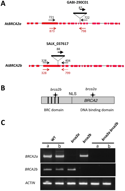
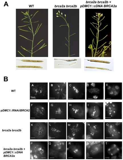
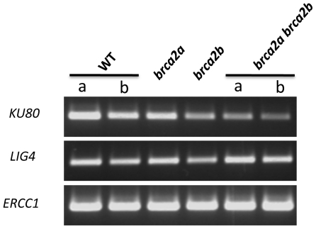
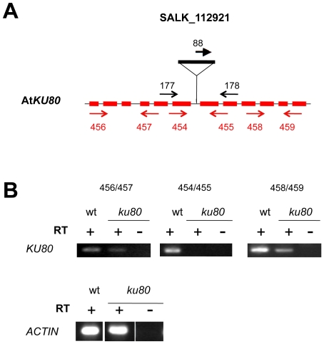
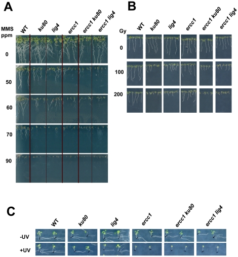
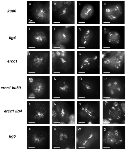
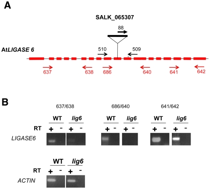
In somatic cells, three major pathways are involved in the repair of DNA double-strand breaks (DBS): Non-Homologous End Joining (NHEJ), Single-Strand Annealing (SSA) and Homologous Recombination (HR). In somatic and meiotic HR, DNA DSB are 5' to 3' resected, producing long 3' single-stranded DNA extensions. Brca2 is essential to load the Rad51 recombinase onto these 3' overhangs. The resulting nucleofilament can thus invade a homologous DNA sequence to copy and restore the original genetic information. In Arabidopsis, the inactivation of Brca2 specifically during meiosis by an RNAi approach results in aberrant chromosome aggregates, chromosomal fragmentation and missegregation leading to a sterility phenotype. We had previously suggested that such chromosomal behaviour could be due to NHEJ. In this study, we show that knock-out plants affected in both BRCA2 genes show the same meiotic phenotype as the RNAi-inactivated plants. Moreover, it is demonstrated that during meiosis, neither NHEJ nor SSA compensate for HR deficiency in BRCA2-inactivated plants. The role of the plant-specific DNA Ligase6 is also excluded. The possible mechanism(s) involved in the formation of these aberrant chromosomal bridges in the absence of HR during meiosis are discussed.


Conflict of interest statement

Competing Interests: The authors have declared that no competing interests exist.

Figures

Figure 1
Figure 1. The brca2 single and double mutants.
(A) Position of the T-DNA insertions in AtBRCA2a and AtBRCA2b. The structure of the AtBRCA2a and AtBRCA2b genes is represented by shaded boxes (exons) and thin lines (introns). The T-DNA insertion position is indicated. Each primer pair used to identify the mutants by PCR are compiled on the diagram in black and primer pairs used for RT-PCR analyses are given in red; their localization is correct but not to scale. (B) Schematically represented Brca2 protein with the position of the BRC repeats and the NLS relative to the T-DNA insertions, as indicated by a star. For convenience, and because they share 94.5% of identity, a single Brca2 protein is represented. (C) RT-PCR analysis of AtBRCA2 transcripts in the single and double brca2 mutants. RNA was extracted from young floral buds of wild-type plants (2 different plants, a and b) as well as of brca2a, brca2b and brca2a brca2b (2 different plants, a and b) mutant plants and was then reverse-transcribed. Double-stranded cDNAs were then PCR-amplified using the primer pairs represented in red in Figure 1A. The constitutive ACTIN gene transcript was used as a control.
Figure 2
Figure 2. Meiotic defects in brca2a brca2b mutant plants and in wild-type Brca2-inactivated plants.
(A) Wild-type and brca2 double mutant plants exhibt no growth defect except for sterility. Chloralhydrate discolored siliques are full of seeds in wild-type plants in comparison with the discolored siliques of the brca2 double mutant plants. (B) Observation of meiocytes by DAPI staining in Brca2-deficient plants, transformed or not with the full length cDNA of AtBRCA2a, and in brca2a brca2b homozygous double mutant plants. (A–E) Different stages of meiosis in the wild-type plants. Meiosis is normal. (A) Prophase I stage, (B) diakinesis, the five bivalents are attached by a chiasma, (C) metaphase I with five aligned bivalents, (D) anaphase I, bivalents segregate into two sets of five univalents, (E) anaphase II, with four groups that contain five chromosomes each after sister chromatid separation. (F–J) Different stages of meiosis in wild-type plants transformed with the pDMC1::RNAi/BRCA2 construct. (F) Prophase I, (G) no normal diakinesis phase (H) metaphase I with condensed and entangled chromosomes, (I) anaphase I, with entangled and stretched chromosomes. (J) Anaphase II, with bridges extending between chromosomes. (K–O) Different stages of meiosis in brca2 double mutant plants. (K) Prophase I, (L) anaphase I, entangled and stretched chromosomes. (M) Metaphase II with entangled chromosomes. (N) anaphase II, fragmentated chromosomes. (O) telophase II with chromosome missegregation. (P–T) Different stages of meiosis in brca2 double mutant plants, transformed with the pDMC1::cDNA AtBRCA2a. Meiosis is restored to normal. (P) Prophase I stage, (Q) diakinesis, (R) metaphase I, (S) anaphase I, (T) anaphase II. Bar 10 µm.
Figure 3
Figure 3. RT-PCR analysis of genes involved in NHEJ and SSA in the single and double brca2 mutants.
RNA was extracted from young floral buds and reverse-transcribed, as described in Figure 1C. Double-stranded cDNAs were PCR-amplified using primer pair 454/455 for AtKU80 (see primer positions in Figure 4 and sequences in Table1), 336/445 for AtLIGIV and 452/453 for AtERCC1 (see Table 1 for sequences). The constitutive ACTIN gene transcript used as a control is presented in Figure 1C.
Figure 4
Figure 4. T-DNA insertion and expression in ku80 mutant.
(A) Position of the T-DNA insertion in AtKU80. The structure of the AtKU80 gene is represented by shaded boxes (exons) and thin lines (introns). The T-DNA insertion position is indicated. Each primer pair used to characterize the mutant by PCR are indicated in black and primer pairs used for RT-PCR analyses are given in red; their localization is correct but not to scale. (B) RT-PCR analysis of AtKU80 transcripts in ku80-/- mutant plants. RNA, extracted from floral buds of wild-type or ku mutant plants was reverse-transcribed. Double-stranded cDNAs were amplified by RT-PCR, performed with three different primer pairs: 5′ or 3′ to the T-DNA and flanking the T-DNA insertion. For primer positions, see above (Figure 4A). The constitutive ACTIN gene was used as a control.
Figure 5
Figure 5. Hypersentivity to MMS, gamma-rays and UV irradiation of nhej, ssa and nhej ssa plants.
Before sowing, all seeds were surface-sterilized. (A) MMS hypersensitivity, 11 days post-germination. Seeds were sown on MS 0.5 agar 1% sucrose supplemented with MMS at various doses. (B) Gamma-irradiation hypersentivity, 7 days post-irradiation. After 48 h at 4°C in darkness, seeds were exposed to various doses of gamma-rays : 0, 100 and 200 grays before being sown on MS 0.5 agar. (C) UV hypersensitivity, 10 days post-irradiation. Seeds were sown in MS 0.5 agar. After 4 days of growth, the plantlets were exposed to UV-C, left in the dark for 3 days to avoid photoreactivation, and then exposed to light.
Figure 6
Figure 6. Observation of meiocytes by DAPI staining in nhej, ssa, nhej ssa and lig6 mutant plants transformed with the pDMC1::RNAi/BRCA2 construct.
Different stages of meiosis were observed in plants transformed with pDMC1::RNAi/BRCA2 in nhej mutant plants, ku80 (A–D) or lig4 (E–H), and in ssa mutant plants, ercc1 (I–L), in nhej ssa double mutant plants, ercc1 ku80 (M–P) or ercc1 lig4 (Q–T) and in lig6 mutant plants (U–X). (A, E, I, M, Q, U) prophase I. (B, F, J, N, R, V) metaphase I. (C, G, K, O, S, W) anaphase I. (D, H, L, P, T, X) anaphase II. Bar 10 µm.
Figure 7
Figure 7. T-DNA insertion and expression in lig6 mutant.
(A) Position of the T-DNA insertion in AtLIG6. The structure of the AtLIG6 gene is represented by shaded boxes (exons) and thin lines (introns). The T-DNA insertion position is indicated. Each primer pair used to identify the mutants by PCR are indicated in black while primer pairs used for RT-PCR analyses are given in red; their localization is correct but not to of scale. (B) RT-PCR analysis of AtLIG6 transcripts in lig6-/- mutant plants. RNA, extracted from floral buds of wild-type or lig6 mutant plants was reverse-transcribed. Double-stranded cDNAs were amplified by RT-PCR, performed with three different primer pairs: 5′ or 3′ to the T-DNAand flanking the T-DNA. The position of each primer is given above (Figure 7A). The constitutive ACTIN gene was used as a control.

References

    1. Daley JM, Palmbos PL, Wu D, Wilson TE. Nonhomologous end joining in yeast. Annu Rev Genet. 2005;39:431–451. - PubMed
    1. Lieber MR. The mechanism of double-strand DNA break repair by the nonhomologous DNA end-joining pathway. Annu Rev Biochem. 2010;79:181–211. - PMC - PubMed
    1. Tamura K, Adachi Y, Chiba K, Oguchi K, Takahashi H. Identification of Ku70 and Ku80 homologues in Arabidopsis thaliana: evidence for a role in the repair of DNA double-strand breaks. Plant J. 2002;29:771–781. - PubMed
    1. Riha K, Watson JM, Parkey J, Shippen DE. Telomere length deregulation and enhanced sensitivity to genotoxic stress in Arabidopsis mutants deficient in Ku70. Embo J. 2002;21:2819–2826. - PMC - PubMed
    1. Friesner J, Britt AB. Ku80- and DNA ligase IV-deficient plants are sensitive to ionizing radiation and defective in T-DNA integration. Plant J. 2003;34:427–440. - PubMed

Publication types