. 2012 Jan 11;134(1):315-23.
doi: 10.1021/ja207148m. Epub 2011 Dec 8.

Evaluation of diverse α/β-backbone patterns for functional α-helix mimicry: analogues of the Bim BH3 domain

Affiliations

Evaluation of diverse α/β-backbone patterns for functional α-helix mimicry: analogues of the Bim BH3 domain

Melissa D Boersma et al. J Am Chem Soc. .

Abstract

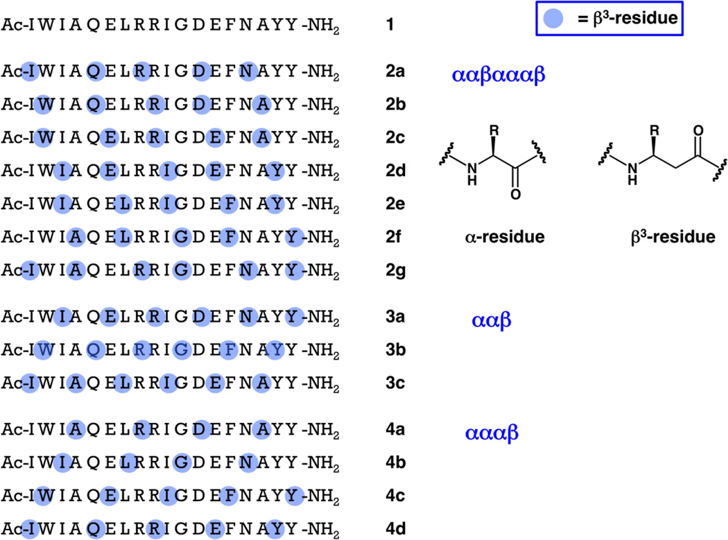
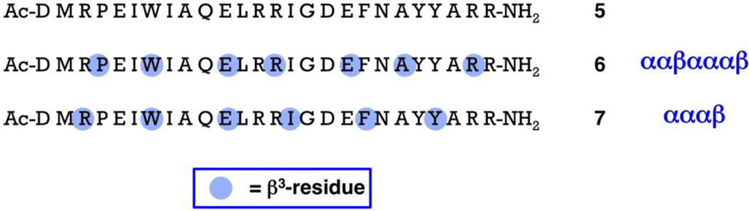
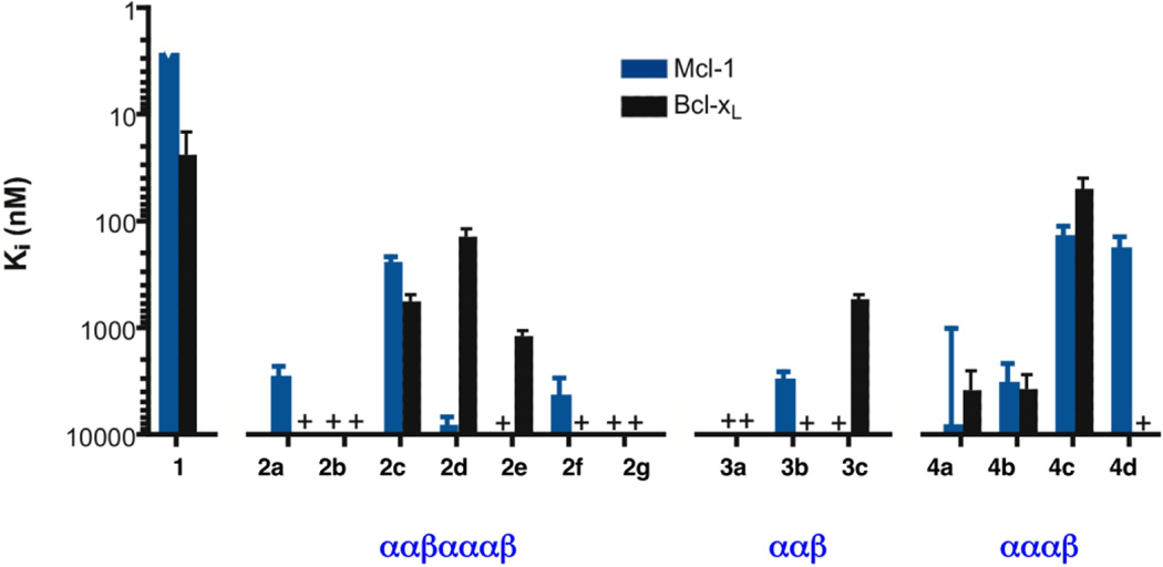
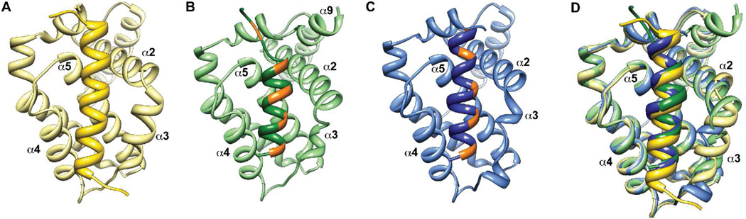
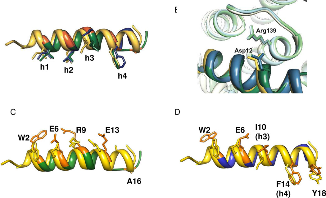
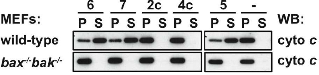
Peptidic oligomers that contain both α- and β-amino acid residues, in regular patterns throughout the backbone, are emerging as structural mimics of α-helix-forming conventional peptides (composed exclusively of α-amino acid residues). Here we describe a comprehensive evaluation of diverse α/β-peptide homologues of the Bim BH3 domain in terms of their ability to bind to the BH3-recognition sites on two partner proteins, Bcl-x(L) and Mcl-1. These proteins are members of the anti-apoptotic Bcl-2 family, and both bind tightly to the Bim BH3 domain itself. All α/β-peptide homologues retain the side-chain sequence of the Bim BH3 domain, but each homologue contains periodic α-residue → β(3)-residue substitutions. Previous work has shown that the ααβαααβ pattern, which aligns the β(3)-residues in a 'stripe' along one side of the helix, can support functional α-helix mimicry, and the results reported here strengthen this conclusion. The present study provides the first evaluation of functional mimicry by ααβ and αααβ patterns, which cause the β(3)-residues to spiral around the helix periphery. We find that the αααβ pattern can support effective mimicry of the Bim BH3 domain, as manifested by the crystal structure of an α/β-peptide bound to Bcl-x(L), affinity for a variety of Bcl-2 family proteins, and induction of apoptotic signaling in mouse embryonic fibroblast extracts. The best αααβ homologue shows substantial protection from proteolytic degradation relative to the Bim BH3 α-peptide.


Figures

Figure 1
Figure 1
Sequences of 18-mer α-peptide 1, derived from the Bim BH3 domain, and 18-mer α/β-peptides of families 2–4. The standard single-letter code is used to designate α-amino acid residues. Blue dots are used to indicate the positions of β3-amino acid residues, which bear the side chain of the α-amino acid residue identified by the single-letter code. Thus, all peptides shown here have the same sequence of side chains, but they have varying backbones (each β3-residue introduces an additional CH2 unit relative to the analogous α-residue).
Figure 2
Figure 2
Sequences of 26-mer α-peptide 5, derived from the Bim BH3 domain, and 26-mer α/β-peptides 6 and 7, which are extensions of 18-mer α/β-peptides 2c and 4c, respectively. The standard single-letter code is used to designate α-amino acid residues. Blue dots are used to indicate the positions of β3-amino acid residues, which bear the side chain of the α-amino acid residue identified by the single-letter code. Thus, peptides 5–7 have the same sequence of side chains, but they have varying backbones (each β3-residue introduces an additional CH2 unit relative to the analogous α-residue).
Figure 3
Figure 3
Graphical summary of inhibition constants (Ki) for binding to Mcl-1 (blue bars) or Bcl-xL (black bars) for 18-mer α-peptide 1 and homologous α/β-peptides in families 2–4, based on competition fluorescence polarization assays (see text for details). Note that the vertical axis is an inverse logarthmic scale, so that taller bars correspond to tighter binding (smaller Ki value, which should correspond to the Kd value).
Figure 4
Figure 4
Crystal structures of α-peptide 5 (A; yellow; PDB 3FDL), α/β-peptide 2c (B; green) and α/β-peptide 4c (C; blue) bound to Bcl-xL. In each case only the backbone is shown. The BH3 domain-derived peptides are indicated by the darker-colored helix at the center of each panel; the lighter-colored portions correspond to Bcl-xL in each panel. For the two α/β-peptides, orange patches indicate the positions of the β3-residues. Both α/β-peptides bind comparably to Bim BH3-derived α-peptide 5 in a groove formed predominantly by helices α2-α5 of Bcl-xL. Part D shows an overlay of the three co-crystal structures, with colors as in parts A–C. Overall the structures are remarkably similar with the exception of the α3 helix of Bcl-xL.
Figure 5
Figure 5
Overlay of α-peptide 5 (yellow), α/β-peptide 2c (green), and α/β-peptide 4c (blue) from the co-crystal structures shown in Figure 4. Part A highlights the positions of the h1–h4 side-chains. Part B highlights the intermolecular salt bridge between an Asp side chain on the outward-facing side of the α- or α/β-peptide and the Arg139 side chain from the Bcl-xL. Parts C and D show pairwise comparisons of α/β-peptide 5 with either α/β-peptide 2c (C) or α/β-peptide 4c (D). These images compare the β3-residue side chains of 2c or 4c with the corresponding α-residue side chains from 5; these side chains sets display a consistent offset for 2c (C) but not for 4c (D) except for the Trp residue, as discussed in the text.
Figure 6
Figure 6
Global analysis of BH3-derived α/β-peptide binding to Bcl-xL (upper panel) or Mcl-1 (lower panel). The numbers across the top of each panel correspond to the sequence positions in the Bim-derived 18-mers introduced in this paper (Figure 1). Five positions characteristic of BH3 domains, four hydrophobic residues (h1–h4) and an Asp residue (D) are indicated. The leftmost column of each panel classifies the α/β-peptides as strong or weak binders to the relevant pro-survival protein. The next column identifies each α- or α/β-peptide, with number designations as in Figure 1. The remaining columns indicate whether the indicated position within the designated α/β-peptide is occupied by a β3-residue (blue box) or by an α-residue (white or green box). The green boxes highlight positions in strong binders that appear to require an α-residue to form a complex with Bcl-xL (upper panel) or with Mcl-1 (lower panel).
Figure 7
Figure 7
Cytochrome c release assay results. Permeabilised wild-type or bax−/−/bak−/− mouse embryonic fibroblasts (MEFs) were treated with 26-mer Bim BH3 α-peptide 5, homologous 26-mer α/β-peptide 6 or 7, or 18-mer α/β-peptide 2c or 4c. All three of the 26-mer oligomers cause cytochrome c release from the pellet fraction (P), which contains mitochondria, into the soluble (S) cytosolic fraction of wild-type MEFs. No release was observed with 18-mer α/β-peptide 2c or 4c, which is consistent with their weaker affinity for pro-survival proteins, relative to 26-mers 5–7. None of the peptides caused any cytochrome c release in bax−/−/bak−/− MEFs.
Figure 8
Figure 8
Circular dichroism data for 26-mer Bim BH3 α-peptide 5 and homologous 26-mer α/β-peptides 6 and 7 (25 µM peptide in 10 mM phosphate buffer, pH 7.5).

References

    1. Jochim AL, Arora PS. Mol. Biosyst. 2009;5:924. - PMC - PubMed
    2. Jochim AL, Arora PS. ACS Chem. Biol. 2010;5:919. - PMC - PubMed
    1. Guarracino DA, Bullock BN, Arora PS. Biopolymers. 2011;95:1. - PMC - PubMed
    2. Guichard G, Huc I. Chem. Comm. 2011;47:5933. - PubMed
    3. Goodman CM, Choi S, Shandler S, DeGrado WF. Nat. Chem. Biol. 2007;3:252. - PMC - PubMed
    4. Hecht S, Huc I. Wenheim: Wiley-VCH; 2007.
    5. Schafmeister CE, Brown ZZ, Gupta S. Acc. Chem. Res. 2008;41:1387. - PubMed
    6. Davis J, Tsou L, Hamilton A. Chem. Soc. Rev. 2006;36:326. - PubMed
    7. Yin H, Hamilton AD. Angew. Chem. Int. Ed. Engl. 2005;44:4130. - PubMed
    1. Werder M, Hauser H, Abele S, Seebach D. Helv. Chim. Acta. 1999;82:1774.
    2. Kritzer JA, Luedtke NW, Harker EA, Schepartz A. J. Am. Chem. Soc. 2005;127:14584. - PMC - PubMed
    3. Stephens OM, Kim S, Welch BD, Hodsdon ME, Kay MS, Schepartz A. J. Am. Chem. Soc. 2005;127:13126. - PMC - PubMed
    4. English EP, Chumanov RS, Gellman SH, Compton TJ. Biol. Chem. 2006;281:2661. - PubMed
    1. Hara T, Durell SR, Myers MC, Appella DH. J. Am. Chem. Soc. 2006;128:1995. - PubMed
    2. Hayashi R, Wang D, Hara T, Iera JA, Durell SR, Appella DH. Bioorg. Med. Chem. 2009;17:7884. - PMC - PubMed
    1. Ernst JT, Kutzki O, Debnath AK, Jiang S, Lu H, Hamilton AD. Angew. Chem. Int. Ed. Engl. 2002;41:278. - PubMed
    2. Kutzki O, Park HS, Ernst JT, Orner BP, Yin H, Hamilton AD. J. Am. Chem. Soc. 2002;124:11838. - PubMed
    3. Ernst JT, Becerril J, Park HS, Yin H, Hamilton AD. Angew. Chem. Int. Ed. Engl. 2003;42:535. - PubMed
    4. Yin H, Lee GI, Sedey KA, Rodriguez JM, Wang HG, Sebti SM, Hamilton AD. J. Am. Chem. Soc. 2005;127:5463. - PubMed
    5. Yin H, Lee GI, Sedey KA, Kutzki O, Park HS, Orner BP, Ernst JT, Wang HG, Sebti SM, Hamilton AD. J. Am. Chem. Soc. 2005;127:10191. - PubMed
    6. Ahn J-M, Han S-Y. Tetrahedron Lett. 2007;28:5343.
    7. Hu X, Sun J, Wang H-G, Manetsch R. J. Am. Chem. Soc. 2008;130:13820. - PMC - PubMed
    8. Shaginian A, Whitby LR, Hong S, Hwang I, Farooqi B, Searcey M, Chen J, Vogt PK, Boger DL. J. Am. Chem. Soc. 2009;131:5564. - PMC - PubMed
    9. Campbell F, Plante JP, Edwards TA, Warriner SL, Wilson A. J. Org. Biomol. Chem. 2010;8:2344. - PubMed
    10. Lee JH, Zhang Q, Jo S, Chai SC, Oh M, Im W, Lu H, Lim H-S. J. Am. Chem. Soc. 2011;133:676. - PMC - PubMed

Publication types

MeSH terms