

Piezoelectric-assisted removal of a benign fibrous histiocytoma of the mandible: an innovative technique for prevention of dentoalveolar nerve injury
- PMID: 22040611
- PMCID: PMC3213176
- DOI: 10.1186/1746-160X-7-20
Piezoelectric-assisted removal of a benign fibrous histiocytoma of the mandible: an innovative technique for prevention of dentoalveolar nerve injury
Abstract
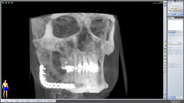
In this article, we present our experience with a piezoelectric-assisted surgical device by resection of a benign fibrous histiocytoma of the mandible.A 41 year-old male was admitted to our hospital because of slowly progressive right buccal swelling. After further radiographic diagnosis surgical removal of the yellowish-white mass was performed. Histologic analysis showed proliferating histiocytic cells with foamy, granular cytoplasm and no signs of malignancy. The tumor was positive for CD68 and vimentin in immunohistochemical staining. Therefore the tumor was diagnosed as primary benign fibrous histiocytoma. This work provides a new treatment device for benign mandibular tumour disease. By using a novel piezoelectric-assisted cutting device, protection of the dentoalveolar nerve could be achieved.
Figures


Similar articles
-
Benign fibrous histiocytoma of the mandible.J Oral Pathol Med. 2005 Mar;34(3):190-2. doi: 10.1111/j.1600-0714.2004.00274.x. J Oral Pathol Med. 2005. PMID: 15689235
-
Clinical and pathologic characteristics and surgical management of benign fibrous histiocytoma of the mandible: a case report.J Oral Maxillofac Surg. 2012 Nov;70(11):2719-23. doi: 10.1016/j.joms.2012.02.004. Epub 2012 May 11. J Oral Maxillofac Surg. 2012. PMID: 22580097 No abstract available.
-
Benign fibrous histiocytoma in the condylar process of the mandible: Case report.Br J Oral Maxillofac Surg. 2008 Jan;46(1):e1-2. doi: 10.1016/j.bjoms.2007.03.020. Epub 2007 Jun 11. Br J Oral Maxillofac Surg. 2008. PMID: 17561319
-
Malignant fibrous histiocytoma of the mandible.Skeletal Radiol. 1996 Jan;25(1):96-9. doi: 10.1007/s002560050043. Skeletal Radiol. 1996. PMID: 8717132 Review.
-
Primary malignant fibrous histiocytoma of the mandible: a case report.East Afr Med J. 1993 Jul;70(7):460-3. East Afr Med J. 1993. PMID: 8293709 Review.
Cited by
-
Benign Spindle Cell Tumour of Mandible and Points of Modification in Reconstruction with Nonvascularised Iliac Crest Graft.J Maxillofac Oral Surg. 2016 Jul;15(Suppl 2):262-5. doi: 10.1007/s12663-015-0775-4. Epub 2015 Mar 24. J Maxillofac Oral Surg. 2016. PMID: 27408449 Free PMC article.
-
Piezoelectric-Assisted Removal of a Mandibular Cementoossifying Fibroma: An Innovative Technique.Case Rep Dent. 2020 Sep 8;2020:8821090. doi: 10.1155/2020/8821090. eCollection 2020. Case Rep Dent. 2020. PMID: 33005458 Free PMC article.
-
Benign Fibrous Histiocytoma: A Rare Case Report and Literature Review.J Maxillofac Oral Surg. 2016 Mar;15(1):116-20. doi: 10.1007/s12663-014-0721-x. Epub 2014 Nov 8. J Maxillofac Oral Surg. 2016. PMID: 26929563 Free PMC article.
-
Immediate loading of one-piece implants in conjunction with a modified technique of inferior alveolar nerve lateralization: 10 years follow-up.Craniomaxillofac Trauma Reconstr. 2014 Mar;7(1):55-62. doi: 10.1055/s-0033-1364198. Epub 2014 Jan 13. Craniomaxillofac Trauma Reconstr. 2014. PMID: 24624258 Free PMC article.
-
Benign fibrous histiocytoma: A rare case involving jaw bone.Contemp Clin Dent. 2015 Sep;6(Suppl 1):S266-8. doi: 10.4103/0976-237X.166828. Contemp Clin Dent. 2015. PMID: 26604585 Free PMC article.
References
-
- Fletcher CDM, Unni KK, Mertens F, World Health Organization; International Agency for Research on Cancer. Pathology and genetics of tumours of soft tissue and bone. Lyon: IARC Press; 2002.
Publication types
MeSH terms
LinkOut - more resources
Full Text Sources
