

Eph/ephrin signaling in epidermal differentiation and disease
- PMID: 22040910
- PMCID: PMC3378995
- DOI: 10.1016/j.semcdb.2011.10.017
Eph/ephrin signaling in epidermal differentiation and disease
Abstract
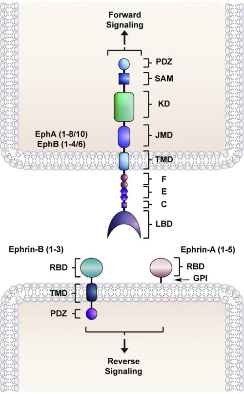
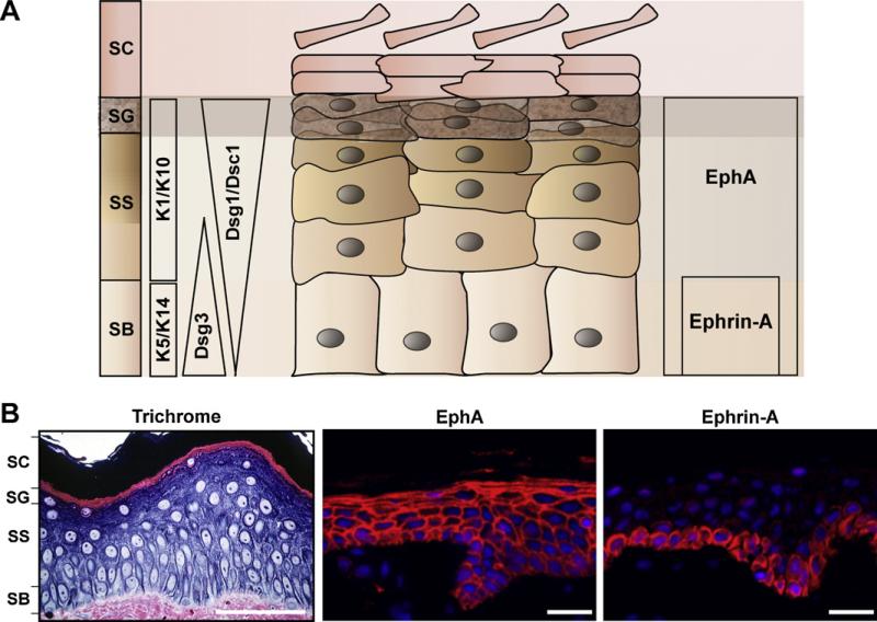
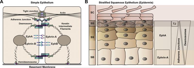
Eph receptor tyrosine kinases mediate cell-cell communication by interacting with ephrin ligands residing on adjacent cell surfaces. In doing so, these juxtamembrane signaling complexes provide important contextual information about the cellular microenvironment that helps orchestrate tissue morphogenesis and maintain homeostasis. Eph/ephrin signaling has been implicated in various aspects of mammalian skin physiology, with several members of this large family of receptor tyrosine kinases and their ligands present in the epidermis, hair follicles, sebaceous glands, and underlying dermis. This review focuses on the emerging role of Eph receptors and ephrins in epidermal keratinocytes where they can modulate proliferation, migration, differentiation, and death. The activation of Eph receptors by ephrins at sites of cell-cell contact also appears to play a key role in the maturation of intercellular junctional complexes as keratinocytes move out of the basal layer and differentiate in the suprabasal layers of this stratified, squamous epithelium. Furthermore, alterations in the epidermal Eph/ephrin axis have been associated with cutaneous malignancy, wound healing defects and inflammatory skin conditions. These collective observations suggest that the Eph/ephrin cell-cell communication pathway may be amenable to therapeutic intervention for the purpose of restoring epidermal tissue homeostasis and integrity in dermatological disorders.
Copyright © 2011 Elsevier Ltd. All rights reserved.
Figures
Similar articles
-
Eph receptor and ephrin function in breast, gut, and skin epithelia.Cell Adh Migr. 2014;8(4):327-38. doi: 10.4161/19336918.2014.970012. Cell Adh Migr. 2014. PMID: 25482622 Free PMC article. Review.
-
The role of Eph receptors and Ephrins in the skin.Int J Dermatol. 2016 Jan;55(1):3-10. doi: 10.1111/ijd.12968. Epub 2015 Oct 24. Int J Dermatol. 2016. PMID: 26498559 Review.
-
Eph/ephrin signaling in morphogenesis, neural development and plasticity.Curr Opin Cell Biol. 2004 Oct;16(5):580-9. doi: 10.1016/j.ceb.2004.07.002. Curr Opin Cell Biol. 2004. PMID: 15363810 Review.
-
Interaxonal Eph-ephrin signaling may mediate sorting of olfactory sensory axons in Manduca sexta.J Neurosci. 2003 Dec 17;23(37):11523-38. doi: 10.1523/JNEUROSCI.23-37-11523.2003. J Neurosci. 2003. PMID: 14684856 Free PMC article.
-
Specific and shared targets of ephrin A signaling in epidermal keratinocytes.J Biol Chem. 2011 Mar 18;286(11):9419-28. doi: 10.1074/jbc.M110.197087. Epub 2010 Dec 30. J Biol Chem. 2011. PMID: 21193391 Free PMC article.
Cited by
-
Single-Cell and Spatial Transcriptomic Analysis of Human Skin Delineates Intercellular Communication and Pathogenic Cells.J Invest Dermatol. 2023 Nov;143(11):2177-2192.e13. doi: 10.1016/j.jid.2023.02.040. Epub 2023 May 2. J Invest Dermatol. 2023. PMID: 37142187 Free PMC article.
-
Single-cell sequencing combined with spatial transcriptomics reveals the characteristics of follicle-targeted inflammation patterns in primary cicatricial alopecia.Cell Biosci. 2025 Jul 16;15(1):102. doi: 10.1186/s13578-025-01447-1. Cell Biosci. 2025. PMID: 40671126 Free PMC article.
-
Complementary expression and repulsive signaling suggest that EphB2 and ephrin-B1 are possibly involved in epithelial boundary formation at the squamocolumnar junction in the rodent stomach.Histochem Cell Biol. 2013 Dec;140(6):659-75. doi: 10.1007/s00418-013-1129-2. Epub 2013 Jul 24. Histochem Cell Biol. 2013. PMID: 23881165
-
Eph signaling is regulated by miRNA-210: Implications for corneal epithelial repair.FASEB J. 2022 Jan;36(1):e22076. doi: 10.1096/fj.202101423R. FASEB J. 2022. PMID: 34856019 Free PMC article.
-
Corneal epithelial biology: Lessons stemming from old to new.Exp Eye Res. 2020 Sep;198:108094. doi: 10.1016/j.exer.2020.108094. Epub 2020 Jul 19. Exp Eye Res. 2020. PMID: 32697979 Free PMC article. Review.
References
-
- Koster MI, Roop DR. Mechanisms regulating epithelial stratification. Annu Rev Cell Dev Biol. 2007;23:93–113. - PubMed
-
- Proksch E, Brandner JM, Jensen JM. The skin: an indispensable barrier. Exp Dermatol. 2008;17(12):1063–72. - PubMed
-
- Green KJ, Simpson CL. Desmosomes: new perspectives on a classic. J Invest Dermatol. 2007;127(11):2499–515. - PubMed
-
- Gumbiner BM. Regulation of cadherin-mediated adhesion in morphogenesis. Nat Rev Mol Cell Biol. 2005;6(8):622–34. - PubMed
Publication types
MeSH terms
Substances
Grants and funding
LinkOut - more resources
Full Text Sources
Other Literature Sources
Miscellaneous
