Review
. 2011 Nov;41(6):1133-69.
doi: 10.1016/j.cvsm.2011.08.004.

Feline coronavirus in multicat environments

Affiliations
Review

Feline coronavirus in multicat environments

Yvonne Drechsler et al. Vet Clin North Am Small Anim Pract. 2011 Nov.

Abstract

Feline infectious peritonitis (FIP), a fatal disease in cats worldwide, is caused by FCoV infection, which commonly occurs in multicat environments. The enteric FCoV, referred to as feline enteric virus (FECV), is considered a mostly benign biotype infecting the gut, whereas the FIP virus biotype is considered the highly pathogenic etiologic agent for FIP. Current laboratory tests are unable to distinguish between virus biotypes of FCoV. FECV is highly contagious and easily spreads in multicat environments; therefore, the challenges to animal shelters are tremendous. This review summarizes interdisciplinary current knowledge in regard to virology, immunology, pathology, diagnostics, and treatment options in the context of multicat environments.


Figures

Fig. 1
Fig. 1
Schematic of the FCoV virion (viral particle). Nucleocapsid proteins coat the RNA genome. The spike, membrane, and envelope proteins are anchored in the bilipid membrane of cell origin.
Fig. 2
Fig. 2
Schematic of the gene organization on the FCoV genome. A cap structure at the 5′ end and the 3′ end poly-adenylated tail are typical structures on an RNA used as message for generating protein within a cell. The entire genome is approximately 29,000 nucleotide bases in length. The overlapping ORFs 1a and 1b encode proteins involved in RNA synthesis required for generating mRNA, the genome, and their negative sense templates. The spike refers to the gene encoding the highly glycosylated spike protein (S), Mem refers to the gene encoding the membrane protein (M), env refers to the gene encoding the envelope protein (E), and nucleocapsid refers to the gene encoding the nucleocapsid protein (N).
Fig. 3
Fig. 3
Peritoneal effusion from a cat with classic wet (or effusive) form of FIP. (A) Characteristic color of peritoneal effusion collected by abdominocentesis. (B) Close view of a plastic bag containing 350 ml of abdominal effusion and large clumps of fibrin. The high viscosity of the effusion due to high protein content can be seen in Video 1.
Fig. 4
Fig. 4
Cat kidneys. (A) Multifocal to coalescing granulomatous inflammation (white, rough appearance) following the superficial blood vessels. (B) Cut section also shows the vascular-oriented distribution.
Fig. 5
Fig. 5
Peritoneal cavity of a cat: intestine, liver, lymph node, spleen, and diaphragm. White-to-yellow soft plaques covering the parietal and visceral peritoneal surfaces (white arrow). The lymph nodes associated with large intestine are enlarged and yellow (black arrow).
Fig. 6
Fig. 6
Spleen from a cat. The capsular surface shows severe fibrinous inflammatory reaction that extends to the omentum. The inflammatory reaction is admixed with copious amounts of fibrin.
Fig. 7
Fig. 7
Cat with wet (effusive) form of FIP presenting moderate abdominal distention due to peritoneal effusion. The abdominal distention is generally not evident in early stages, and may require imaging techniques to be confirmed.
Fig. 8
Fig. 8
Lateral thoracic radiograph image of a cat with pleural effusion due to FIP.
Fig. 9
Fig. 9
(A) Anterior uveitis typically seen in noneffusive cases of FIP. Mild iridal neovascularization (rubeosis iridis) and hyphema are evident in the anterior chamber of the right eye (OD). (B) Fibrin formation, hypopyon, and evidence of mild diapedesis are suggestive of blood–ocular barrier breakdown associated with mild anterior uveitis. (C) Severe iritis, with rubeosis iridis, aqueous flare, hypopyon, and keratitic precipitates. These precipitates, known as “mutton-fat” precipitates, are suggestive of a chronic granulomatous disease process.
Fig. 10
Fig. 10
Kidney. Superficial renal venules. Necrotic tubular epithelial cells (white arrows) with severe interstitial pyogranulomatous inflammation. The small venule (black arrow) contains an intravascular fibrin thrombus and with moderate mural vascular necrosis (hematoxylin-eosin, original magnification ×20).
Fig. 11
Fig. 11
Spinal cord. (A) There is severe pyogranulomatous inflammation that is most intense around the blood (hematoxylin-eosin, original magnification ×60). (B) The vessel wall is stained in brown and shows thickening of the wall by moderate to severe smooth muscle hyperplasia (smooth muscle actin with peroxidase stain, original magnification ×60).
Fig. 12
Fig. 12
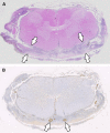
Spinal cord. (A) Subgross cross-section with marked thickening of the meninges due to pyogranulomatous inflammation (between white arrows). (B) Immunohistochemistry for smooth muscle actin indicates marked medial thickening of the small or medium-size vessels due to smooth muscle hyperplasia (white arrows).
Fig. 13
Fig. 13
Brain, lateral ventricle from a cat with FIP (FCoV immunohistochemistry stain). Macrophages within the lesion have intense cytoplasmic staining (gold-brownish color), confirming the presence of viral antigen (monoclonal antibody 1:400, original magnification ×60).

References

    1. Rohrbach B.W., Legendre A.M., Baldwin C.A., et al. Epidemiology of feline infectious peritonitis among cats examined at veterinary medical teaching hospitals. J Am Vet Med Assoc. 2001;218(7):1111–1115. - PubMed
    1. Cave T.A., Thompson H., Reid S.W., et al. Kitten mortality in the United Kingdom: a retrospective analysis of 274 histopathological examinations (1986 to 2000) Vet Rec. 2002;151(17):497–501. - PubMed
    1. Decaro N., Buonavoglia C. An update on canine coronaviruses: viral evolution and pathobiology. Vet Microbiol. 2008;132(3-4):221–234. - PMC - PubMed
    1. Garwes D.J. Transmissible gastroenteritis. Vet Rec. 1988;122(19):462–463. - PubMed
    1. Perlman S., Dandekar A.A. Immunopathogenesis of coronavirus infections: implications for SARS. Nat Rev Immunol. 2005;5(12):917–927. - PMC - PubMed

MeSH terms