

Amygdalar, hippocampal, and thalamic volumes in youth at high risk for development of bipolar disorder
- PMID: 22041532
- PMCID: PMC3225692
- DOI: 10.1016/j.pscychresns.2011.03.006
Amygdalar, hippocampal, and thalamic volumes in youth at high risk for development of bipolar disorder
Abstract
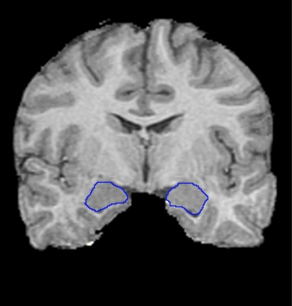
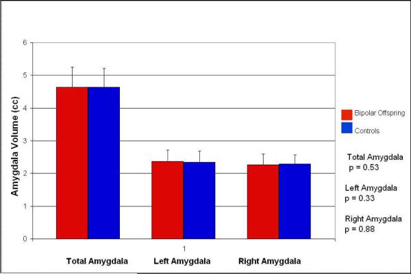
Children of parents with bipolar disorder (BD), especially those with attention deficit hyperactivity disorder (ADHD) and symptoms of depression or mania, are at significantly high risk for developing BD. As we have previously shown amygdalar reductions in pediatric BD, the current study examined amygdalar volumes in offspring of parents (BD offspring) who have not yet developed a full manic episode. Youth participating in the study included 22 BD offspring and 22 healthy controls of comparable age, gender, handedness, and IQ. Subjects had no history of a manic episode, but met criteria for ADHD and moderate mood symptoms. MRI was performed on a 3T GE scanner, using a 3D volumetric spoiled gradient echo series. Amygdalae were manually traced using BrainImage Java software on positionally normalized brain stacks. Bipolar offspring had similar amygdalar volumes compared to the control group. Exploratory analyses yielded no differences in hippocampal or thalamic volumes. Bipolar offspring do not show decreased amygdalar volume, possibly because these abnormalities occur after more prolonged illness rather than as a preexisting risk factor. Longitudinal studies are needed to determine whether amygdalar volumes change during and after the development of BD.
2011 Elsevier Ireland Ltd. All rights reserved.
Figures
References
-
- Adolphs R, Baron-Cohen S, Tranel D. Impaired recognition of social emotions following amygdala damage. Journal of Cognitive Neuroscience. 2002;14:1264–74. - PubMed
-
- Adolphs R, Tranel D. Impaired judgments of sadness but not happiness following bilateral amygdala damage. Journal of Cognitive Neuroscience. 2004;16:453–462. - PubMed
-
- Altshuler LL, Bartzokis G, Grieder T, Curran J, Jimenez T, Leight K, Wilkins J, Gerner R, Mintz J. An MRI study of temporal lobe structures in men with bipolar disorder or schizophrenia. Biological Psychiatry. 2000;48:147–162. - PubMed
-
- Altshuler LL, Bartzokis G, Grieder T, Curran J, Mintz J. Amygdala enlargement in bipolar disorder and hippocampal reduction in schizophrenia: an MRI study demonstrating neuroanatomic specificity. Archives of General Psychiatry. 1998;55:663–664. - PubMed
-
- Altshuler LL, Bookheimer SY, Townsend J, Proenza MA, Eisenberger N, Sabb F, Mintz J, Cohen MS. Blunted activation in orbitofrontal cortex during mania: a functional magnetic resonance imaging study. Biological Psychiatry. 2005;58:763–769. - PubMed
Publication types
MeSH terms
Grants and funding
LinkOut - more resources
Full Text Sources
Medical
Research Materials
Miscellaneous
