. 2011 Nov 8;108(45):18342-7.
doi: 10.1073/pnas.1106550108. Epub 2011 Oct 31.

CD47-signal regulatory protein-α (SIRPα) interactions form a barrier for antibody-mediated tumor cell destruction

Affiliations

CD47-signal regulatory protein-α (SIRPα) interactions form a barrier for antibody-mediated tumor cell destruction

Xi Wen Zhao et al. Proc Natl Acad Sci U S A. .

Abstract

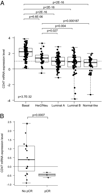
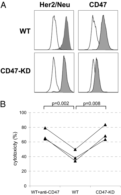
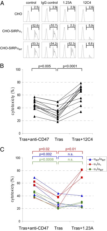
Monoclonal antibodies are among the most promising therapeutic agents for treating cancer. Therapeutic cancer antibodies bind to tumor cells, turning them into targets for immune-mediated destruction. We show here that this antibody-mediated killing of tumor cells is limited by a mechanism involving the interaction between tumor cell-expressed CD47 and the inhibitory receptor signal regulatory protein-α (SIRPα) on myeloid cells. Mice that lack the SIRPα cytoplasmic tail, and hence its inhibitory signaling, display increased antibody-mediated elimination of melanoma cells in vivo. Moreover, interference with CD47-SIRPα interactions by CD47 knockdown or by antagonistic antibodies against CD47 or SIRPα significantly enhances the in vitro killing of trastuzumab-opsonized Her2/Neu-positive breast cancer cells by phagocytes. Finally, the response to trastuzumab therapy in breast cancer patients appears correlated to cancer cell CD47 expression. These findings demonstrate that CD47-SIRPα interactions participate in a homeostatic mechanism that restricts antibody-mediated killing of tumor cells. This provides a rational basis for targeting CD47-SIRPα interactions, using for instance the antagonistic antibodies against human SIRPα described herein, to potentiate the clinical effects of cancer therapeutic antibodies.


Conflict of interest statement

The authors declare no conflict of interest.

Figures

Fig. 1.
Fig. 1.
SIRPα signaling limits antibody-mediated destruction of melanoma cells in vivo. (A) CD47 expression on B16F10 mouse melanoma cells as demonstrated by flow cytometry using anti-mouse CD47 antibody (Miap301) and phycoerythrin-labeled anti-mouse IgG (filled histogram). The open histogram represents the isotype control. (B) Comparable outgrowth of B16 melanoma in wild-type and SIRPα-mutant mice in the absence of therapeutic antibody. Wild-type and SIRPα-mutant mice were injected i.v. with 1.5 × 10⁵ B16F10 tumor cells. After 21 d, mice were killed, lungs were excised and photographed (representative examples are shown), and tumor loads were determined and expressed as the sum of the following scores: metastases less than 1 mm were scored as 1; metastases between 1 and 2 mm were scored as 3; and metastases larger than 2 mm were scored as 10. Measurements from individual mice are shown, with means indicated by bars, and statistical differences between groups (n = 10) were determined by ANOVA. Note that comparable tumor loads occur in wild-type (34.7 ± 9.5) (mean ± SEM) and SIRPα-mutant mice (35.9 ± 5.2). Data are from one representative experiment out of three. (C) Enhanced antibody-mediated clearance of B16 melanoma cells in SIRPα-mutant mice. Wild-type and SIRPα-mutant mice were challenged i.v. with 1.5 × 10⁵ B16F10 tumor cells and, where indicated, with a suboptimal dose of 10 μg of TA99 antibody (or PBS as control) on days 0, 2, and 4. After 21 d, mice were killed and analyzed as in B. Measurements from individual mice are shown, with means indicated by bars, and statistical differences between groups (n = 8) were determined by ANOVA. Note the black nodules of melanoma lung metastases in B and C. Note in the graph in C that TA99 antibody treatment resulted only in a minimal nonsignificant reduction in tumor cell outgrowth in wild-type animals [47.9 ± 9.4 (mean ± SEM) in PBS-treated mice compared with 29.0 ± 7.8 in TA99-treated mice], but tumor formation was essentially absent in SIRPα-mutant animals treated with TA99 antibody (4.5 ± 1.0). Data are from one representative experiment out of three.
Fig. 2.
Fig. 2.
CD47 mRNA expression in breast cancer. (A) Correlation with molecular subtypes: basal, Her2/Neu-positive, luminal A, luminal B, and normal-like (Institut Paoli-Calmettes series; n = 353). Log2-transformed expression levels in tumors are reported as box plots relative to expression in normal breast (NB; horizontal solid line). Overexpression (ratio T:NB ≥2; horizontal dashed line) of CD47 was found in 63% of tumors. Note that the poor-prognosis subtypes (i.e., basal and Her2/Neu+) have the highest CD47 expression levels. Differences in expression levels between the five subtypes were tested for significance using one-way ANOVA, and between two subtypes using Student's t test. (B) Correlation with pathological response to trastuzumab plus vinorelbine treatment [public data set (29); n = 22]. Log2-transformed expression levels in tumors are reported as box plots relative to median expression in all samples (median; horizontal solid line). Note that patients with a pathological complete response (pCR; n = 3) have significantly lower CD47 expression than patients with an incomplete response (no pCR; n = 19).
Fig. 3.
Fig. 3.
Interference with CD47–SIRPα interactions using blocking anti-CD47 antibody B6H12 potentiates trastuzumab-mediated ADCC of neutrophils toward Her2/Neu-positive SKBR-3 breast cancer cells. (A) Flow cytometric analysis of Her2/Neu and CD47 surface expression on SKBR-3 breast cancer cells (filled histograms), using trastuzumab and B6H12 mAb, respectively, against CD47. Isotype controls are shown in the open histograms. (B) ADCC of neutrophils against trastuzumab-opsonized SKBR-3 cells (E:T ratio, 50:1) in the absence or presence of B6H12 anti-CD47 F(ab′)2. Shown is a representative example. Results are expressed as means ± SD of triplicate measurements, and statistical differences were shown by Student's t test. Note that anti-CD47 F(ab′)2 fragments do not affect cytotoxicity alone, but do synergize with trastuzumab. (C and D) Blocking CD47–SIRPα interactions using anti-CD47 F(ab′)2 enhances the ADCC of neutrophils against trastuzumab-opsonized SKBR-3 cells at different E:T ratios (C) and trastuzumab concentrations (D). Shown is a representative experiment out of three. (E) The effects of anti-CD47 F(ab′)2 on ADCC toward trastuzumab-opsonized SKBR-3 cells using neutrophils from different donors in multiple independent experiments (n = 53). For clarity, only the values in the presence of trastuzumab ± anti-CD47 F(ab′)2 are shown, with the matched values of the two conditions for each donor connected by lines. Killing in the absence of trastuzumab ± anti-CD47 F(ab′)2 was always below 5%. P values of statistically significant differences, as determined by Student's t test, are indicated.
Fig. 4.
Fig. 4.
Knockdown of CD47 in SKBR-3 breast cancer target cells enhances trastuzumab-dependent neutrophil-mediated ADCC. (A) Flow cytometric analysis for Her2/Neu and CD47 surface expression in SKBR-3 cells transfected with empty vector (control) or CD47 shRNA (CD47-KD). Note that CD47 expression is strongly decreased in the CD47-KD cells (mean fluorescence intensity (MFI) = 358 in CD47-KD cells vs. MFI = 4.187 in control), but Her2/Neu levels are unaltered (MFI = 18.638 in CD47-KD cells and MFI = 18.993 in control). (B) Neutrophil-mediated ADCC using control and CD47-KD SKBR-3 cells opsonized with trastuzumab in three independent experiments with three different effector cell donors. Note that a similar level of enhancement occurs with anti-CD47 F(ab′)2-mediated blocking and CD47 knockdown. P values of statistically significant differences, as determined by Student's t test, are indicated.
Fig. 5.
Fig. 5.
Monoclonal antibodies against SIRPα that block CD47–SIRPα interactions enhance ADCC. (A) CD47-coated fluorescent bead binding to CHO cells expressing empty vector (i.e., “CHO”), SIRPα1, or SIRPαBIT. The 12C4 and 1.23A mAbs (but not isotype IgG1 control mAb) block the binding of CD47 beads to either both SIRPα1- and SIRPαBIT-expressing CHO cells (12C4) or only to SIRPα1-expressing CHO cells. The proportion (in %) of cells binding CD47 beads is indicated in the upper right of each panel. Shown is one representative experiment out of three. (B) Enhancing effect of 12C4 mAb on ADCC toward trastuzumab-opsonized SKBR-3 cells using neutrophils from (n = 12) individuals in four independent experiments. (C) Enhancing effect of 1.23A mAb on ADCC toward trastuzumab-opsonized SKBR-3 cells using neutrophils from (n = 9) individuals with different SIRPα genotypes (α11 or αBITBIT homozygotes or α1BIT heterozygotes) in three independent experiments. P values of statistically significant differences, as determined by Student's t test, are indicated. n.s., nonsignificant.

References

    1. Glennie MJ, van de Winkel JG. Renaissance of cancer therapeutic antibodies. Drug Discov Today. 2003;8:503–510. - PubMed
    1. Oldham RK, Dillman RO. Monoclonal antibodies in cancer therapy: 25 years of progress. J Clin Oncol. 2008;26:1774–1777. - PubMed
    1. Strome SE, Sausville EA, Mann D. A mechanistic perspective of monoclonal antibodies in cancer therapy beyond target-related effects. Oncologist. 2007;12:1084–1095. - PubMed
    1. Clynes RA, Towers TL, Presta LG, Ravetch JV. Inhibitory Fc receptors modulate in vivo cytoxicity against tumor targets. Nat Med. 2000;6:443–446. - PubMed
    1. Barok M, et al. Trastuzumab causes antibody-dependent cellular cytotoxicity-mediated growth inhibition of submacroscopic JIMT-1 breast cancer xenografts despite intrinsic drug resistance. Mol Cancer Ther. 2007;6:2065–2072. - PubMed

MeSH terms