. 2011 Nov 15;108(46):18825-30.
doi: 10.1073/pnas.1116449108. Epub 2011 Oct 31.

Gating charge interactions with the S1 segment during activation of a Na+ channel voltage sensor

Affiliations

Gating charge interactions with the S1 segment during activation of a Na+ channel voltage sensor

Paul G DeCaen et al. Proc Natl Acad Sci U S A. .

Abstract

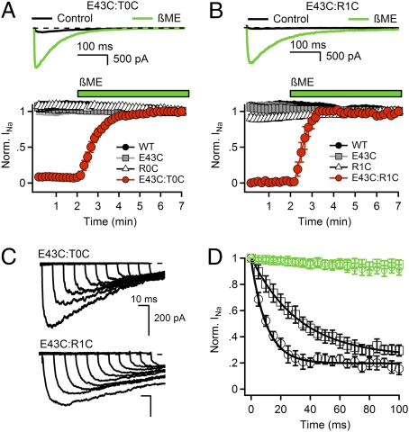
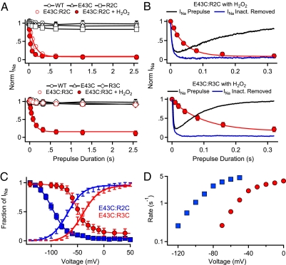
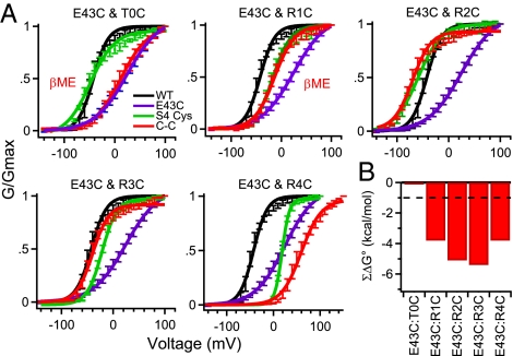
Voltage-gated Na(+) channels initiate action potentials during electrical signaling in excitable cells. Opening and closing of the pore of voltage-gated ion channels are mechanically linked to voltage-driven outward movement of the positively charged S4 transmembrane segment in their voltage sensors. Disulfide locking of cysteine residues substituted for the outermost T0 and R1 gating-charge positions and a conserved negative charge (E43) at the extracellular end of the S1 segment of the bacterial Na(+) channel NaChBac detects molecular interactions that stabilize the resting state of the voltage sensor and define its conformation. Upon depolarization, the more inward gating charges R2 and R3 engage in these molecular interactions as the S4 segment moves outward to its intermediate and activated states. The R4 gating charge does not disulfide-lock with E43, suggesting an outer limit to its transmembrane movement. These molecular interactions reveal how the S4 gating charges are stabilized in the resting state and how their outward movement is catalyzed by interaction with negatively charged residues to effect pore opening and initiate electrical signaling.


Conflict of interest statement

The authors declare no conflict of interest.

Figures

Fig. 1.
Fig. 1.
Disulfide locking of the voltage sensors in the resting state of E43C:T0C and E43C:R1C channels. (A and B) Upper: INa from the first pulse in control conditions (black) and last pulse in the presence of reducing agent (green) elicited by a 0.1-Hz train of 500-ms depolarizations to 0 mV from a holding potential of −140 mV. Lower: Effects of 10 mM βME on the mean normalized peak currents (±SEM) recorded during trains (n = 6). (C) Rate of relocking in the resting state. E43C:T0C and E43C:R1C channels were unlocked by a 5-s pulse to −180 mV in the presence of 2 mM βME. Cells were then returned to βME-free solution for 5 min, depolarized to −120 mV for 0, 10, 20, 30, 40, 50, 60, 70, 80, 90, and 100 ms, and INa was measured by a 500-ms test pulse to 0 mV. (D) Rates of relocking of the voltage sensors in the resting state at −120 mV. βME (2 mM) was removed (black) or retained (green) during this phase of the experiment. Mean normalized peak currents are plotted vs. time at −120 mV (±SEM, n = 4). E43C:T0C (black circles), τ = 10.3 ms; E43C:R1C (black squares), τ = 32.5 ms; E43C:T0C + βME (green circles); E43C:R1C + βME (green squares).
Fig. 2.
Fig. 2.
Disulfide locking of E43:R2C and E43C:R3C requires activation. Mean normalized peak currents during trains of 500-ms depolarizations to V = V1/2 + 40 mV from a holding potential of −140 mV. (AC) E43C:R2C and single mutants as indicated. (A) Disulfide locking of E43C:R2C. After 1 min, pulsing was stopped for 5 min to test for recovery of INa. (B) Effect of H2O2 on disulfide locking of E43C:R2C. Disulfide locking in the presence of 2 mM H2O2 for 2 min at −140 mV without pulsing followed by 2 min with a train of 500-ms depolarizations. (C) Effect of reduction with βME on E43C:R2C. After 2 min of stimulation, cells were exposed to 10 mM βME, and stimulation was continued (n = 5). Current Insets are the first pulse in the presence of H2O2 (black), the last pulse in the presence of H2O2 (gray), and the last pulse in the presence of reducing agent (green). (DF) E43C:R2C and single mutants as indicated. (D) Effect of 2 mM H2O2 on disulfide locking of E43C:R2C. (E) Requirement for depolarization for disulfide locking of E43C:R2C. Cells were stimulated with 500-ms depolarizations for 1 min, exposed to H2O2 for 1 min at −140 mV without pulsing, and pulsing was resumed. (F) Reversal of disulfide locking with 10 mM βME. Cells were stimulated with 500-ms depolarizations for 2 min, H2O2 was added for 3 min with continued stimulation, H2O2 was washed out for 3 min, and 10 mM βME was added for 5 min (n = 4). Mean ± SEM. Current Insets are the first pulse in the presence of H2O2 (black), the last pulse in the presence of H2O2 (blue), and the last pulse in the presence of reducing agent (green).
Fig. 3.
Fig. 3.
Time course and voltage dependence of disulfide locking of E43C:R2C and E43C:R3C channels. (A) E43C:R2C (Upper) and E43C:R3C channels (Lower) were unlocked by a 5-s prepulse to −170 mV. Cells were then depolarized by prepulses to V1/2 + 40 mV of the indicated durations. After 5 s at −140 mV, disulfide-locked channels were assayed with a 100-ms test pulse to V1/2 + 40 mV (WT, 0 mV; E43C, 60 mV; R2C, −20 mV; E43C:R2C, −30 mV; E43C:R3C, 0 mV). Peak test pulse currents were normalized to the test pulse current in the absence of a prepulse. Mean values (±SEM) were plotted vs. prepulse duration (n = 6). (B) Comparison of the rate of disulfide locking and channel activation (blue line, after subtraction of the exponential effect of inactivation) for E43C:R2C and E43C:R3C in 2 mM H2O2. (C) The voltage dependence of channel activation (lines with error bars) and of disulfide locking (symbols) measured in the presence of 2 mM H2O2. To measure the voltage dependence of disulfide locking of E43C:R2C (n = 9), channels were unlocked by a 5-s pulse to −170 mV, then depolarized with 500-ms prepulses to the indicated potentials. After 5 s at −140 mV, the number of channels locked during the prepulse was assessed with a 100-ms test pulse to 0 mV. Because H2O2-induced disulfide locking of E43C:R3C channels cannot be reversed by hyperpolarization, disulfide locking could only be tested at one potential for each cell (n = 5–6 for each potential). (D) Rate of disulfide locking as a function of membrane potential estimated from the data in C at potentials where the fraction of channels locked was between 0.1 and 0.9 as ln[(Fraction of INa)]/(−0.5 s).
Fig. 4.
Fig. 4.
Mutant cycle analysis of cysteine pairs. (A) Conductance–voltage relationships for WT, double-mutant channels, and the E43C and gating charge single-mutant channels. Conductance–voltage relationships for double mutants E43C:T0C and E43C:R1C were measured in the presence of 10 mM βME. Conductance–voltage relationships were calculated from peak INa elicited by 100-ms depolarizations to the indicated potentials (n ≥ 9; ±SEM) as described in SI Methods. (B) Coupling energy (ΣΔG°) for each residue pair as described in SI Methods. Mean ± SEM.
Fig. 5.
Fig. 5.
Transmembrane view of disulfide-locked Rosetta Membrane structural models of the voltage-sensing domain of NaChBac. Segments S1 through S4 colored individually and labeled. Side chains of cysteine mutants of E43, T0, R1, R2, and R3 are shown in space-filling representation and colored yellow. Cβ atoms of gating-charge-carrying arginines (blue; labeled R1 through R4) in S4 and D60 and E70 (red) in S2 shown as spheres. Models were generated with the Rosetta Membrane modeling system (SI Methods). This figure was generated using Chimera (43).

References

    1. Hille B. Ion Channels of Excitable Membranes. 3rd Ed. Sunderland, MA: Sinauer; 2001.
    1. Ito M, et al. The voltage-gated Na+ channel NaVBP has a role in motility, chemotaxis, and pH homeostasis of an alkaliphilic Bacillus. Proc Natl Acad Sci USA. 2004;101:10566–10571. - PMC - PubMed
    1. Yu FH, Catterall WA. The VGL-chanome: A protein superfamily specialized for electrical signaling and ionic homeostasis. Sci STKE. 2004;2004:re15. - PubMed
    1. Yi BA, Jan LY. Taking apart the gating of voltage-gated K+ channels. Neuron. 2000;27:423–425. - PubMed
    1. Yellen G. The voltage-gated potassium channels and their relatives. Nature. 2002;419:35–42. - PubMed

Publication types