. 2011 Dec 6;108(49):E1339-48.
doi: 10.1073/pnas.1102349108. Epub 2011 Oct 31.

FGF2 gene transfer restores hippocampal functions in mouse models of Alzheimer's disease and has therapeutic implications for neurocognitive disorders

Affiliations

FGF2 gene transfer restores hippocampal functions in mouse models of Alzheimer's disease and has therapeutic implications for neurocognitive disorders

Tomomi Kiyota et al. Proc Natl Acad Sci U S A. .

Erratum in

  • Proc Natl Acad Sci U S A. 2011 Dec 27;108(52):21282

Abstract

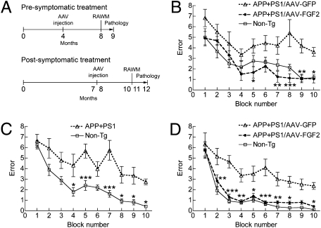
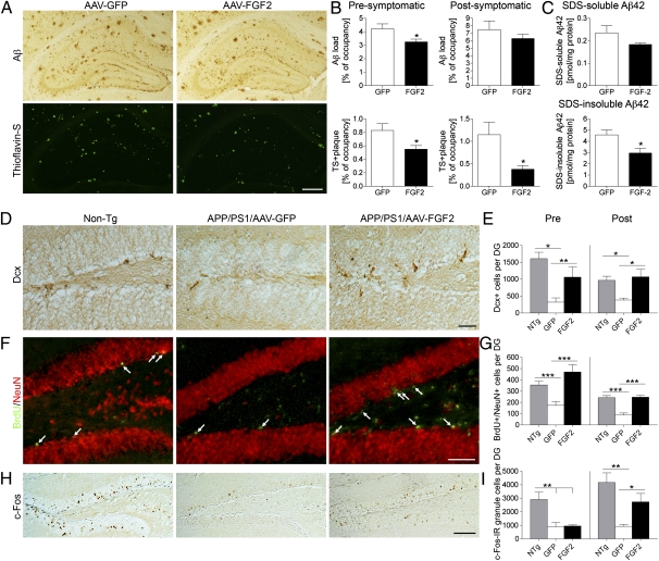
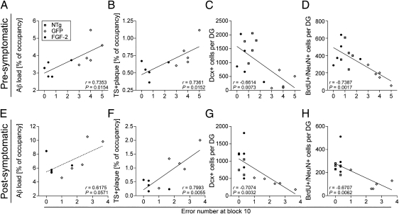
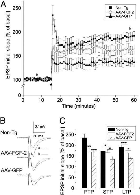
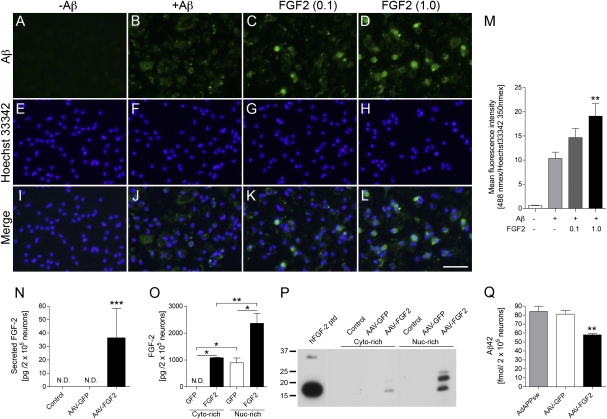
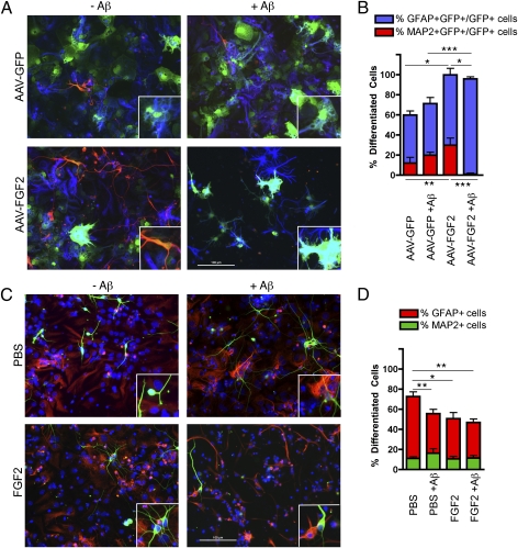
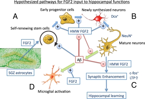
The adult hippocampus plays a central role in memory formation, synaptic plasticity, and neurogenesis. The subgranular zone of the dentate gyrus contains neural progenitor cells with self-renewal and multilineage potency. Transgene expression of familial Alzheimer's disease-linked mutants of β-amyloid precursor protein (APP) and presenilin-1 leads to a significant inhibition of neurogenesis, which is potentially linked to age-dependent memory loss. To investigate the effect of neurogenesis on cognitive function in a relevant disease model, FGF2 gene is delivered bilaterally to the hippocampi of APP+presenilin-1 bigenic mice via an adenoassociated virus serotype 2/1 hybrid (AAV2/1-FGF2). Animals injected with AAV2/1-FGF2 at a pre- or postsymptomatic stage show significantly improved spatial learning in the radial arm water maze test. A neuropathological investigation demonstrates that AAV2/1-FGF2 injection enhances the number of doublecortin, BrdU/NeuN, and c-fos-positive cells in the dentate gyrus, and the clearance of fibrillar amyloid-β peptide (Aβ) in the hippocampus. AAV2/1-FGF2 injection also enhances long-term potentiation in another APP mouse model (J20) compared with control AAV2/1-GFP-injected littermates. An in vitro study confirmed the enhanced neurogenesis of mouse neural stem cells by direct AAV2/1-FGF2 infection in an Aβ oligomer-sensitive manner. Further, FGF2 enhances Aβ phagocytosis in primary cultured microglia, and reduces Aβ production from primary cultured neurons after AAV2/1-FGF2 infection. Thus, our data indicate that virus-mediated FGF2 gene delivery has potential as an alternative therapy of Alzheimer's disease and possibly other neurocognitive disorders.


Conflict of interest statement

The authors declare no conflict of interest.

Figures

Fig. 1.
Fig. 1.
Gene delivery of FGF2 improves memory function of APP+PS1 mice. (A) Experimental designs for pre- and postsymptomatic AAV injections into APP+PS1 mice. APP+PS1 mice received bilateral hippocampal injections of AAV2/1-GFP or AAV2/1-FGF2 at 4 mo or 7 to 8 mo of age, and were tested by a RAWM task at 8 mo or 10 to 11 mo of age (n = 8 per group) for pre- or postsymptomatic study, respectively. Non-Tg mice served as positive controls for the spatial learning task. (B) APP+PS1 mice received hippocampal injections of AAV2/1-GFP or AAV2/1-FGF2 at 4 mo of age (presymptomatic group), and were posttested in the RAWM task at 8 mo of age (n = 8 per group). (C) A preconditioning test of a RAWM task for non-Tg (n = 12) and untreated APP+PS1 (n = 24) groups was conducted at 7 mo of age. Both learning memory and recall were impaired in the APP+PS1 mice at this point. (D) APP+PS1 mice received hippocampal injections of AAV2/1-GFP or AAV2/1-FGF2 after the preconditioning test (postsymptomatic group), and were posttested in the RAWM task at 10 to 11 mo of age (n = 12 per group). *P < 0.05, **P < 0.01, or ***P < 0.001 vs. AAV2/1-GFP group (or vs. non-Tg, C) as determined by two-way repeated-measures ANOVA and Bonferroni posttest, respectively.
Fig. 2.
Fig. 2.
Gene delivery of FGF2 reduces Aβ deposition and enhances neurogenesis in the hippocampal region of the APP+PS1 mouse brain. (A) Representative images of Aβ staining in the hippocampus of APP+PS1 mice in the postsymptomatic treatment. Sections were counterstained by TS for compact plaques. (Scale bar: 500 μm.) (B) Quantification of total Aβ load and TS+ areas in hippocampal regions. Error bars represent SEM (n = 5 per group, five sections per brain). (C) The levels of SDS-soluble Aβ42 and SDS-insoluble Aβ42 in the brain (postsymptomatic group) were measured by human Aβ42-specific ELISA (n = 5). *P < 0.05 vs. AAV2/1-GFP group, as determined by Student t test. (D) Dcx staining in the dentate gyrus of non-Tg or APP+PS1 mice in the postsymptomatic treatment. (Scale bar: 50 μm.) (E) Quantification of Dcx+ cells in the SGZ. (F) Immunofluorescence of BrdU (green) and NeuN (red) staining in the SGZ after postsymptomatic treatment. Arrows indicate BrdU+/NeuN+ double-positive cells. (Scale bar: 100 μm.) (G) Quantification of BrdU+/NeuN+ cells in the SGZ. Mice were intraperitoneally injected with BrdU 3 wk before the euthanasia. (H) c-fos staining in the dentate gyrus after the postsymptomatic treatment. (Scale bars: 200 μm.) (I) Quantification of c-fos+ cells in the SGZ. *P < 0.05, **P < 0.01, or ***P < 0.001 as determined by one-way ANOVA and Newman–Keuls posttest, respectively (n = 5 per group, 10 sections per brain). Error bars represent SEM.
Fig. 3.
Fig. 3.
Significant correlations between spatial learning and β-amyloidosis or neurogenesis. Pearson correlation analysis shows a significant correlation (r value) between the error number at block 10 of the RAWM test and total Aβ load (A, but not E), TS+ plaque load (B and F), Dcx+ cell count (C and G), and BrdU+/NeuN+ cell count (D and H) in presymptomatic (AD) and postsymptomatic treatments (EH).
Fig. 4.
Fig. 4.
Chronic expression of FGF2 attenuates the deficit in LTP in PDAPP (J20) mice. (A) The time course and average magnitude of LTP recorded in the CA1 dendritic field (stratum radium) of hippocampal slices prepared from 5-mo-old J20 mice injected with AAV2/1-GFP (10 slices from four mice), AAV2/1-FGF2 (nine slices from four mice) and non-Tg controls (13 slices from five mice). The graph plots the initial slope of the field EPSPs evoked in response to constant current stimulation. HFS (100 Hz) was delivered at the time indicated by an arrow. Each point in this graph represents an average of three consecutive sweeps. (B) Representative field EPSPs taken 7 min before (dotted line) and 43 min after (solid line) HFS in different experimental groups as indicated. (C) A summary bar graph showing the magnitudes of PTP, STP, and LTP recorded from mice injected with AAV2/1-GFP, AAV2/1-FGF2, or non-Tg controls. *P < 0.05, **P < 0.01, or ***P < 0.001 as determined by one-way ANOVA and Tukey post-hoc test, respectively. Error bars represent SEM.
Fig. 5.
Fig. 5.
FGF2 enhances Aβ phagocytosis in primary microglia and inhibits Aβ production from primary neurons. (A–M) Primary mouse microglia were incubated with or without fibrillar Aβ42 (±Aβ) and recombinant murine FGF2 protein (0.1 or 1.0 ng/mL) for 1 h, followed by immunofluorescence for Aβ (green; AD) and cell nuclei (blue; EH). Merged captured images are also shown (IL). (Scale bar: 50 μm.) (M) Quantification of Aβ fluorescence intensity (excitation/emission wavelengths, 488 nm/519 nm) normalized by Hoechst 33342 intensity (excitation/emission wavelengths, 350 nm/461 nm). **P < 0.01 vs. +Aβ group as determined by one-way ANOVA and Newman–Keuls posttest, respectively (n = 6 per group). Error bars represent SEM. (NQ) Mouse primary cultured neurons (200,000 cells per well in 48-well plate) were infected with or without AAV2/1-GFP (4 × 10¹⁰ VP per 500 μL medium), or AAV2/1-FGF2 (4 × 10¹⁰ VP per 500 μL medium) for 3 d. (N) FGF2 secretion from the neurons (undetectable from uninfected or AAV2/1-GFP–infected cells; 36.50 ± 22.02 pg per 200,000 neurons from AAV2/1-FGF2–infected cells). (O and P) AAV2/1-infected neurons were fractionated into cytoplasm (Cyto)-rich and nuclear (Nuc)-rich fractions, and subjected to FGF2 ELISA (N) or immunoblotting (P). hFGF2 ptd; 18-kDa human FGF2 recombinant peptide. (Q) Mouse primary cultured neurons (200,000 cells per well in a 48-well plate) were infected with adenovirus expressing Swedish familial AD APP695 mutant (AdAPPsw) for 1 h. After washing and one-overnight incubation, the neurons were infected with or without AAV2/1-GFP or AAV2/1-FGF2 for 3 d. Tissue culture media was subjected to Aβ42 ELISA. *P < 0.05, **P < 0.01, or ***P < 0.001 as determined by one-way ANOVA and Newman–Keuls posttest, respectively (n = 3 per group). Error bars represent SEM.
Fig. 6.
Fig. 6.
AAV2/1-FGF2 enhances differentiation of mouse neural stem cells. (A) Dissociated mouse neuronal stem cells infected with AAV2/1-GFP (1.5 × 10¹⁰ VP) or AAV2/1-FGF2 + AAV2/1-GFP (1.5 × 10¹⁰ plus 1.5 × 10⁹ VP) were incubated with or without 1 μM Aβ42 oligomer for 7 d, followed by immunofluorescence for MAP-2 (neuronal marker; red), GFAP (astrocytic marker; blue), and GFP (all AAV-infected cells; green). (B) Quantification of percentage of MAP-2+/GFP+ and GFAP+/GFP+ cells over the total number of GFP+ cells (n = 10 fields of view per group). (C) Uninfected dissociated mouse neuronal stem cells were incubated with or without 1 μM Aβ42 oligomer, in the presence or absence of FGF2 (2 ng/mL) for 7 d, followed by immunofluorescence of MAP-2 (green), GFAP (red), and Hoechst 33342 (blue). (D) Quantification of percentage of MAP-2+ and GFAP+ cells over the total number of Hoechst 33342-expressing cells (n = 10 fields of view per group). (A and C) Original magnification of 200×; inset, 2× magnification of the field. (B and D) Columns represent mean ± SEM; P < 0.05, **P < 0.01, or ***P < 0.001 between two groups as determined by ANOVA and Tukey post-hoc test.
Fig. P1.
Fig. P1.
Schematic diagram of FGF2, Aβ, and hippocampal functions. (A) The proliferation of self-renewing neuronal stem cells in the SGZ of the dentate gyrus is enhanced by a secreted LMW isoform of FGF2 mainly from astrocytes in the SGZ, and potentially by direct stimulation with Aβ. AAV-FGF2 infection in neuronal stem cells expresses intracellular and HMW FGF2 and enhances neuronal differentiation via a distinct mechanism from that of the LMW FGF2. (B) Neuronal maturation is enhanced by HMW FGF2 expression in the neuronal stem cells and is inhibited by Aβ. Neurons also produce Aβ, which is partially inhibited by the expression of HMW FGF2. (C) HMW FGF2 enhances hippocampal neuronal c-fos gene expression and CA1 LTP, and confers the synaptic inhibition by Aβ. Enhanced neuronal plasticity may facilitate learning and memory. (D) LMW FGF2 activates microglia and enhances Aβ uptake, leading to the reduction of Aβ plaques in the brain.

References

    1. Martin JB. Molecular basis of the neurodegenerative disorders. N Engl J Med. 1999;340:1970–1980. - PubMed
    1. Forman MS, Trojanowski JQ, Lee VM. Neurodegenerative diseases: A decade of discoveries paves the way for therapeutic breakthroughs. Nat Med. 2004;10:1055–1063. - PubMed
    1. Selkoe DJ. Alzheimer's disease is a synaptic failure. Science. 2002;298:789–791. - PubMed
    1. Mattson MP. Pathways towards and away from Alzheimer's disease. Nature. 2004;430:631–639. - PMC - PubMed
    1. Blennow K, de Leon MJ, Zetterberg H. Alzheimer's disease. Lancet. 2006;368:387–403. - PubMed

Publication types

MeSH terms

LinkOut - more resources