

2-(3-{1-Carboxy-5-[(6-[18F]fluoro-pyridine-3-carbonyl)-amino]-pentyl}-ureido)-pentanedioic acid, [18F]DCFPyL, a PSMA-based PET imaging agent for prostate cancer
- PMID: 22042970
- PMCID: PMC3243762
- DOI: 10.1158/1078-0432.CCR-11-1357
2-(3-{1-Carboxy-5-[(6-[18F]fluoro-pyridine-3-carbonyl)-amino]-pentyl}-ureido)-pentanedioic acid, [18F]DCFPyL, a PSMA-based PET imaging agent for prostate cancer
Abstract
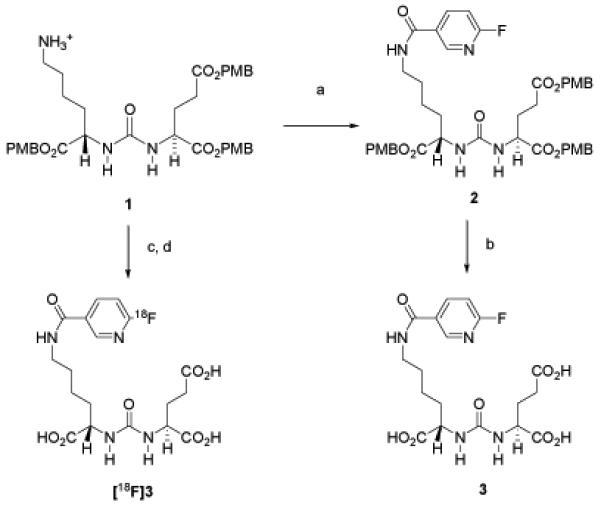
Purpose: We have synthesized and evaluated in vivo 2-(3-{1-carboxy-5-[(6-[(18)F]fluoro-pyridine-3-carbonyl)-amino]-pentyl}-ureido)-pentanedioic acid, [(18)F]DCFPyL, as a potential imaging agent for the prostate-specific membrane antigen (PSMA). PSMA is upregulated in prostate cancer epithelia and in the neovasculature of most solid tumors.
Experimental design: [(18)F]DCFPyL was synthesized in two steps from the p-methoxybenzyl (PMB) protected lys-C(O)-glu urea precursor using 6-[(18)F]fluoronicotinic acid tetrafluorophenyl ester ([(18)F]F-Py-TFP) for introduction of (18)F. Radiochemical synthesis was followed by biodistribution and imaging with PET in immunocompromised mice using isogenic PSMA PC3 PIP and PSMA- PC3 flu xenograft models. Human radiation dosimetry estimates were calculated using OLINDA/EXM 1.0.
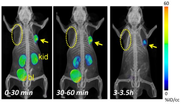
Results: DCFPyL displays a K(i) value of 1.1 ± 0.1 nmol/L for PSMA. [(18)F]DCFPyL was produced in radiochemical yields of 36%-53% (decay corrected) and specific radioactivities of 340-480 Ci/mmol (12.6-17.8 GBq/μmol, n = 3). In an immunocompromised mouse model [(18)F]DCFPyL clearly delineated PSMA+ PC3 PIP prostate tumor xenografts on imaging with PET. At 2 hours postinjection, 39.4 ± 5.4 percent injected dose per gram of tissue (%ID/g) was evident within the PSMA+ PC3 PIP tumor, with a ratio of 358:1 of uptake within PSMA+ PC3 PIP to PSMA- PC3 flu tumor placed in the opposite flank. At or after 1 hour postinjection, minimal nontarget tissue uptake of [(18)F]DCFPyL was observed. The bladder wall is the dose-limiting organ.
Conclusions: These data suggest [(18)F]DCFPyL as a viable, new positron-emitting imaging agent for PSMA-expressing tissues.
©2011 AACR.
Figures
References
-
- Jemal A, Siegel R, Xu J, Ward E. Cancer statistics, 2010. CA: a cancer journal for clinicians. 2010;60:277–300. - PubMed
-
- Ross JS, Sheehan CE, Fisher HA, Kaufman RP, Jr., Kaur P, Gray K, et al. Correlation of primary tumor prostate-specific membrane antigen expression with disease recurrence in prostate cancer. Clin Cancer Res. 2003;9:6357–62. - PubMed
-
- Perner S, Hofer MD, Kim R, Shah RB, Li H, Moller P, et al. Prostate-specific membrane antigen expression as a predictor of prostate cancer progression. Human pathology. 2007;38:696–701. - PubMed
-
- Horoszewicz JS, Kawinski E, Murphy GP. Monoclonal antibodies to a new antigenic marker in epithelial prostatic cells and serum of prostatic cancer patients. Anticancer research. 1987;7:927–35. - PubMed
-
- Smith-Jones PM, Vallabhajosula S, Navarro V, Bastidas D, Goldsmith SJ, Bander NH. Radiolabeled monoclonal antibodies specific to the extracellular domain of prostate-specific membrane antigen: preclinical studies in nude mice bearing LNCaP human prostate tumor. J Nucl Med. 2003;44:610–7. - PubMed
Publication types
MeSH terms
Substances
Grants and funding
LinkOut - more resources
Full Text Sources
Other Literature Sources
Medical
Miscellaneous
