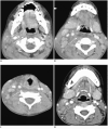
CT analysis of retropharyngeal abnormality in Kawasaki disease
- PMID: 22043152
- PMCID: PMC3194774
- DOI: 10.3348/kjr.2011.12.6.700
CT analysis of retropharyngeal abnormality in Kawasaki disease
Abstract
Objective: To retrospectively compare the imaging characteristics of retropharyngeal density and associated findings for Kawasaki disease with those for non-Kawasaki disease, and identify the distinguishing features which aid the CT diagnosis of Kawasaki disease with retropharyngeal low density.
Materials and methods: Among the enhanced neck CT performed in children less than 8-years old with clinical presentation of fever and cervical lymphadenopathy over a 6-year period, only cases with retropharyngeal low density (RLD) were included in this study. The 56 cases of RLD were divided into two groups; group A included cases diagnosed as Kawasaki disease (n = 34) and group B included cases diagnosed as non-Kawasaki disease (n = 22). We evaluated the CT features including the thickness of RLD and its extent into the deep neck spaces, as well as soft tissue change in the adjacent structure. We also scored the extent of RLD into the deep neck spaces and the soft tissue changes in the adjacent structure.
Results: The thickness of RLD was greater in group A than in group B (group A, 6.0 ± 2.1; group B, 4.6 ± 1.5, p = 0.01). The score of the RLD extent into the deep neck spaces was significantly greater in group A than in group B (group A, 2.3 ± 1.3; group B, 0.8 ± 1.0, p < 0.01). Also, the score of the adjacent soft tissue changes was greater in group A than in group B (group A, 2.0 ± 1.1; group B, 1.0 ± 1.0, p < 0.01).
Conclusion: If children present with fever and cervical lymphadenopathy that display retropharyngeal low density with extension into more deep neck spaces as well as changes in more adjacent soft tissue, the possibility of Kawasaki disease should be considered.
Keywords: CT; Kawasaki disease; Retropharyngeal edema.
Figures


References
-
- Davis WL, Harnsberger HR, Smoker WR, Watanabe AS. Retropharyngeal space: evaluation of normal anatomy and diseases with CT and MR imaging. Radiology. 1990;174:59–64. - PubMed
-
- Elden LM, Grundfast KM, Vezina G. Accuracy and usefulness of radiographic assessment of cervical neck infections in children. J Otolaryngol. 2001;30:82–89. - PubMed
-
- Gross M, Eliashar R, Attal P, Sichel JY. Radiology quiz case 2: Kawasaki disease (KD) mimicking a retropharyngeal abscess. Arch Otolaryngol Head Neck Surg. 2001;127:1507, 1508–1509. - PubMed
-
- Hester TO, Harris JP, Kenny JF, Albernaz MS. Retropharyngeal cellulitis: a manifestation of Kawasaki disease in children. Otolaryngol Head Neck Surg. 1993;109:1030–1033. - PubMed
-
- Homicz MR, Carvalho D, Kearns DB, Edmonds J. An atypical presentation of Kawasaki disease resembling a retropharyngeal abscess. Int J Pediatr Otorhinolaryngol. 2000;54:45–49. - PubMed
Publication types
MeSH terms
LinkOut - more resources
Full Text Sources
Medical

