

Cardiosphere-derived cells improve function in the infarcted rat heart for at least 16 weeks--an MRI study
- PMID: 22043289
- PMCID: PMC3197153
- DOI: 10.1371/journal.pone.0025669
Cardiosphere-derived cells improve function in the infarcted rat heart for at least 16 weeks--an MRI study
Abstract
Aims: Endogenous cardiac progenitor cells, expanded from explants via cardiosphere formation, present a promising cell source to prevent heart failure following myocardial infarction. Here we used cine-magnetic resonance imaging (MRI) to track administered cardiosphere-derived cells (CDCs) and to measure changes in cardiac function over four months in the infarcted rat heart.
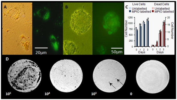
Methods and results: CDCs, cultured from neonatal rat heart, comprised a heterogeneous population including cells expressing the mesenchymal markers CD90 and CD105, the stem cell marker c-kit and the pluripotency markers Sox2, Oct3/4 and Klf-4. CDCs (2 × 10(6)) expressing green fluorescent protein (GFP+) were labelled with fluorescent micron-sized particles of iron oxide (MPIO). Labelled cells were administered to the infarcted rat hearts (n = 7) by intramyocardial injection immediately following reperfusion, then by systemic infusion (4 × 10(6)) 2 days later. A control group (n = 7) was administered cell medium. MR hypointensities caused by the MPIOs were detected at all times and GFP+ cells containing MPIO particles were identified in tissue slices at 16 weeks. At two days after infarction, cardiac function was similar between groups. By 6 weeks, ejection fractions in control hearts had significantly decreased (47 ± 2%), but this was not evident in CDC-treated hearts (56 ± 3%). The significantly higher ejection fractions in the CDC-treated group were maintained for a further 10 weeks. In addition, CDC-treated rat hearts had significantly increased capillary density in the peri-infarct region and lower infarct sizes. MPIO-labelled cells also expressed cardiac troponin I, von Willebrand factor and smooth muscle actin, suggesting their differentiation along the cardiomyocyte lineage and the formation of new blood vessels.
Conclusions: CDCs were retained in the infarcted rat heart for 16 weeks and improved cardiac function.
Conflict of interest statement
Figures


References
-
- Martin-Rendon E, Brunskill SJ, Hyde CJ, Stanworth SJ, Mathur A, et al. Autologous bone marrow stem cells to treat acute myocardial infarction: a systematic review. Eur Heart J. 2008;29:1807–1818. - PubMed
-
- Beltrami AP, Barlucchi L, Torella D, Baker M, Limana F, et al. Adult cardiac stem cells are multipotent and support myocardial regeneration. Cell. 2003;114:763–776. - PubMed
-
- Messina E, De Angelis L, Frati G, Morrone S, Chimenti S, et al. Isolation and expansion of adult cardiac stem cells from human and murine heart. Circ Res. 2004;95:911–921. - PubMed
-
- Smith RR, Barile L, Cho HC, Leppo MK, Hare JM, et al. Regenerative potential of cardiosphere-derived cells expanded from percutaneous endomyocardial biopsy specimens. Circulation. 2007;115:896–908. - PubMed
Publication types
MeSH terms
Substances
Grants and funding
LinkOut - more resources
Full Text Sources
Other Literature Sources
Medical
Research Materials
