. 2011;21(2):93-122.
doi: 10.1615/jlongtermeffmedimplants.v21.i2.10.

Short-term and long-term effects of orthopedic biodegradable implants

Affiliations

Short-term and long-term effects of orthopedic biodegradable implants

Ami R Amini et al. J Long Term Eff Med Implants. 2011.

Abstract

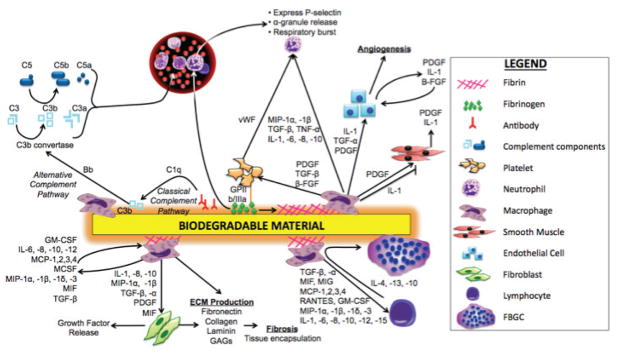
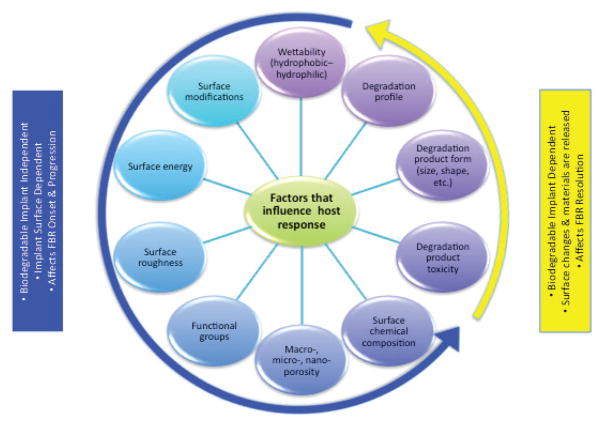
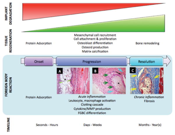
Presently, orthopedic and oral/maxillofacial implants represent a combined $2.8 billion market, a figure expected to experience significant and continued growth. Although traditional permanent implants have been proved clinically efficacious, they are also associated with several drawbacks, including secondary revision and removal surgeries. Non-permanent, biodegradable implants offer a promising alternative for patients, as they provide temporary support and degrade at a rate matching tissue formation, and thus, eliminate the need for secondary surgeries. These implants have been in clinical use for nearly 25 years, competing directly with, or maybe even exceeding, the performance of permanent implants. The initial implantation of biodegradable materials, as with permanent materials, mounts an acute host inflammatory response. Over time, the implant degradation profile and possible degradation product toxicity mediate long-term biodegradable implant-induced inflammation. However, unlike permanent implants, this inflammation is likely to cease once the material disappears. Implant-mediated inflammation is a critical determinant for implant success. Thus, for the development of a proactive biodegradable implant that has the ability to promote optimal bone regeneration and minimal detrimental inflammation, a thorough understanding of short- and long-term inflammatory events is required. Here, we discuss an array of biodegradable orthopedic implants, their associated short- and long- term inflammatory effects, and methods to mediate these inflammatory events.


Figures

FIGURE 1
FIGURE 1
Biodegradable orthopedic implants. (A) Screws, plates, rods and tacks, commonly utilized for fracture fixation and meniscal repair (reprinted from Ciccone et al. 2001). (B) Plates and cages for spinal repair. (C) Porous scaffolds for bone tissue engineering [reprinted from Journal of the American Academy of Orthopaedic Surgeons, vol. 9, no. 5, pp. 280–8, Ciccone WJ, Motz C, Bentley C, Tasto JP. Bioabsorbable implants in orthopaedics: new developments and clinical applications. Copyright (2001), with permission from Elsevier].
FIGURE 2
FIGURE 2
Schematic illustration of biodegradable material-induced inflammation.,,,,
FIGURE 3
FIGURE 3
Histological analysis of a subcutaneous mouse model of nylon mesh biomaterial implantation was performed by analysis of tissue sections of the implant and lesion area at 4 weeks post-implantation (reprinted from Higgins et al.2009). (A, B) Immunohistochemical staining of macrophages and FBGCs in implant tissue sections. Tissue sections were stained for (A) F4/80 and (B) CD11b. (C, D) Immunohistochemical staining of (C) B220 for B cells and (D) CD3 for T cells in implant tissue sections. Arrows indicate stained lymphocytes. (E) The distances between stained CD3, B220 cells or FBGCs and the nearest implant surface. (F) Adhesion cell density. (G) Percentage of macrophage fusion [reprinted from the American Journal of Pathology, vol. 175, no. 1, pp. 161–70, Higgins D, Basaraba R, Hohnbaum A, Lee E, Grainger D, Gonzalez-Juarrero M. Localized immunosuppressive environment in the foreign body response to implanted biomaterials. Copyright (2009), with permission from Elsevier].
FIGURE 4
FIGURE 4
Stage-dependent implant-mediated inflammation. Factors that mediate short-term inflammatory responses to biodegradable implants are independent of their biodegradability characteristics, and instead dependent on trauma-induced inflammation and implant surface characteristics. Long-term inflammation induced by these implants, however, is largely affected by their biodegradation.
FIGURE 5
FIGURE 5
(A) A foreign-body reaction to biodegradable fixation devices made of polyglycolide resulted in a sinus discharging polymeric debris. Observed sinus 11 weeks postoperative on the lateral side of the left ankle of a 43-yr-old man with a bimalleolar fracture that was treated by open reduction and internal fixation using polyglycolide screws. (B) Anteroposterior radiograph of osteolytic lesions (asterisks) at the implant tracks in a 45-yr-old woman with a trimalleolar fracture of the right ankle that was treated by open reduction and internal fixation using polyglycolide screws, 9 weeks postoperative. (C) The characteristic histopathological picture of a non-specific foreign-body reaction to biodegradable implants. Polymeric particles of various sizes (asterisks) are seen surrounded by foreign-body giant cells. Masson-Goldner stain, original magnification 350x [reprinted from Biomaterials, vol. 21, no. 24, pp. 2615–21, Bostman O, PihlajamaÅnki H. Clinical biocompatibility of biodegradable orthopaedic implants for internal f xation: a review. Copyright (2000), with permission from Elsevier].
FIGURE 6
FIGURE 6
Comparison of non-specific walling-off phenomenon resulting from permanent and biodegradable implants. Schematic drawing of the operated femur (A,B). The distal end of the femur shows the polymer implant (1) and metallic implant (2) in the drill hole (A), and the distal half of the femur shows the bioabsorbable (1) and metallic pin (2) placement in the intramedullary canal in the anteroposterior view (B). In the histologic specimens, a walling-off response was observed for both bioabsorbable and metallic pins (C–F). Fibrous tissue and bone trabecula outlining the PDS pin at 3 weeks (C). A bone rim is seen outlining the metallic wire at 12 weeks (D). A fibrous tissue zone surrounds the PGA pin at 24 weeks (E). Thin bone trabecula outlining the PLLA pin at 52 weeks (F). * Implant channel; black arrows, bone; arrowheads, fibrous tissue; white arrow, PDS particles. Masson-Goldner trichrome staining. (reprinted from Pihlajamäki et al. 2010.)
FIGURE 7
FIGURE 7
Biodegradable orthopedic implant-induced inflammation, and the parallel tissue regeneration and wound-healing paradigm. Characteristic histopathological picture of a non-specific foreign-body reaction to biodegradable implants include (A) acute inflammation marked by neutrophils (green arrows); (B) chronic inflammation marked by multinucleated giant cells (dark green arrows), fibrosis (light blue arrows), and mixed inflammatory cells (black arrow); and (C) fibrosis around the implant (yellow arrows) stained with Masson’s trichrome stain (collagen is stained blue). These images were obtained from samples generated by implanting PVA hydrogel/PLGA microsphere composites (marked by *) into the subcutaneous tissue of rats after (A) 3, (B) 30, and (C) 60 days. Bar = 100 μm [reprinted from the Journal of Diabetes Science and Technology, vol. 2, no. 6, pp. 1003–15, Onuki Y, Bhardwaj U, Papadimitrakopoulos F, Burgess D. A review of the biocompatibility of implantable devices: current challenges to overcome foreign body response. Copyright (20089), with permission from Diabetes Technology Society].
FIGURE 8
FIGURE 8
Pharmacodynamic changes in representative tissue sections after subcutaneous implantation of PLGA microsphere/PVA hydrogel composites (HC) containing dexamethasone at (A) day 7 and (B) day 30 post-implantation. (C) Untreated normal tissue. Hematoxylin and eosin stain, HC = hydrogel composite [reprinted from the Journal of Diabetes Science and Technology, vol. 2, no. 6, pp. 1003–15, Onuki Y, Bhardwaj U, Papadimitrakopoulos F, Burgess D. A review of the biocompatibility of implantable devices: current challenges to overcome foreign body response. Copyright (20089), with permission from Diabetes Technology Society].

References

    1. Reichert WM, Ratner BD, Anderson J, Coury A, Hoffman AS, Laurencin CT, Tirrell D. 2010 Panel on the Biomaterials Grand Challenges. J Biomed Mater Res A. 2011 Feb;96(2):275–87. - PMC - PubMed
    1. Mittal R, Morley J, Dinopoulos H, Drakoulakis E, Vermani E, Giannoudis P. Use of bioresorbable implants for stabilisation of distal radius fractures: the United Kingdom patients’ perspective. Injury. 2005 Feb;36(2):333–8. - PubMed
    1. Kulkarni RK, Pani KC, Neuman C, Leonard F. Polylactic acid for surgical implants. Arch Surg. 1966 Nov;93(5):839–43. - PubMed
    1. Rokkanen P, Böstman O, Vainionpää S, Makela EA, Hirvensalo E, Partio EK, Vihtonen K, Pätiälä H, Törmälä P. Absorbable devices in the fixation of fractures. J Trauma. 1996 Mar;40(3 Suppl):S123–7. - PubMed
    1. Rokkanen P, Böstman O, Hirvensalo E, Mäkelä E, Partio E, Pätiälä H, Vainionpää SI, Vihtonen K, Törmälä P. Bioabsorbable fixation in orthopaedic surgery and traumatology. Biomaterials. 2000 Dec;21(24):2607–13. - PubMed