Review
. 2012 Jan;135(1):9-18.
doi: 10.1111/j.1365-2567.2011.03515.x.

Structural and biophysical determinants of αβ T-cell antigen recognition

Affiliations
Review

Structural and biophysical determinants of αβ T-cell antigen recognition

John S Bridgeman et al. Immunology. 2012 Jan.

Abstract

The molecular rules that govern MHC restriction, and allow T-cells to differentiate between peptides derived from healthy cells and those from diseased cells, remain poorly understood. Here we provide an overview of the structural constraints that enable the T-cell receptor (TCR) to discriminate between self and non-self peptides, and summarize studies that have attempted to correlate the biophysical parameters of TCR/peptide-major histocompatibility complex (pMHC) binding with T-cell activation. We further review how the antigenic origin of peptide epitopes affects TCR binding parameters and the 'quality' of a T-cell response. Understanding the principles that govern pMHC recognition by T-cells will unlock pathways to the rational development of immunotherapeutic approaches for the treatment of infectious disease, cancer and autoimmunity.


Figures

Figure 1
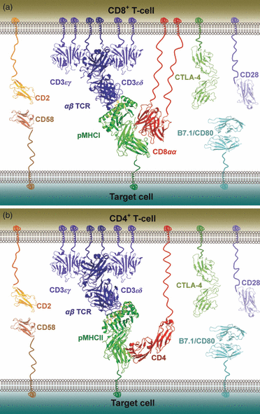
Figure 1
Schematic representation of the molecules involved at the interface between a T-cell and a peptide–MHC (pMHC)-presenting cell during antigen recognition. The co-stimulatory molecule CD28 is shown in mauve and the co-inhibitory molecule CTLA-4 is shown in dark green on the T-cell surface; their cognate ligand, B7.1/CD80, is shown in cyan on the target cell surface. The adhesion molecule CD2 is shown in orange on the T-cell surface and its receptor, CD58, is shown in brown on the target cell surface. The CD3 co-signalling complex is shown in blue. (a) CD8+ T-cell, with the αβ T-cell receptor (αβ TCR) shown in blue, pMHCI shown in green, and the CD8αα co-receptor shown in red. Note that the vast majority of CD8+ T-cells express the CD8αβ heterodimer, but the structure of human CD8αβ is not available for representation. (b) CD4+ T-cell, with the αβ TCR shown in blue, pMHCII shown in green, and the CD4 co-receptor shown in red.
Figure 2
Figure 2
Overview of T-cell receptor (TCR) binding to peptide–MHC (pMHC). The pMHC molecule is shown from above. (a) Surface representation of the footprint of an archetypal αβ TCR (AS01), with the α-chain in green and the β-chain in blue. This diagonal orientation, with the TCR α-chain over the MHCI α2 domain or the MHCII β-chain, and the TCR β-chain over the MHCI α1 domain or the MHCII α chain, has been observed in all TCR/pMHC co-complexes. (b) Positions of the CDR loops during antigen recognition. The CDR1 (α chain in red, β chain in yellow) and CDR3 (α chain in blue, β chain in orange) loops are positioned centrally along the axis of the MHC peptide-binding groove, contacting both the peptide and the MHC surface. The CDR2 (α chain in green, β chain in cyan) loops are positioned such that they contact mainly the MHC surface and make limited, or no, contact with the peptide.
Figure 3
Figure 3
Representation of typical T-cell receptor (TCR) binding affinities. This schematic representation illustrates the mean binding affinities for each type of TCR/peptide–MHC (pMHC) interaction shown. (a) Self-specific TCRs binding to either pMHCI or pMHCII. (b) Non-self-specific TCRs binding to either pMHCI or pMHCII. (c) pMHCI-specific TCRs. (d) pMHCII-specific TCRs. The difference in mean affinity between TCRs specific for self and non-self antigens supports the notion that strongly reactive self-specific T-cells are deleted in the thymus. The weaker mean binding affinity of pMHCII-specific TCRs compared with pMHCI-specific TCRs may reflect the different biological functions of CD4+ and CD8+ T-cells during adaptive immune responses.

References

    1. Gao GF, Rao Z, Bell JI. Molecular coordination of αβ T-cell receptors and coreceptors CD8 and CD4 in their recognition of peptide-MHC ligands. Trends Immunol. 2002;23:408–13. - PubMed
    1. Janeway CA., Jr The T cell receptor as a multicomponent signalling machine: CD4/CD8 coreceptors and CD45 in T cell activation. Annu Rev Immunol. 1992;10:645–74. - PubMed
    1. Barber EK, Dasgupta JD, Schlossman SF, Trevillyan JM, Rudd CE. The CD4 and CD8 antigens are coupled to a protein-tyrosine kinase (p56lck) that phosphorylates the CD3 complex. Proc Natl Acad Sci U S A. 1989;86:3277–81. - PMC - PubMed
    1. Veillette A, Bookman MA, Horak EM, Bolen JB. The CD4 and CD8 T cell surface antigens are associated with the internal membrane tyrosine-protein kinase p56lck. Cell. 1988;55:301–8. - PubMed
    1. Arcaro A, Gregoire C, Bakker TR, et al. CD8β endows CD8 with efficient coreceptor function by coupling T cell receptor/CD3 to raft-associated CD8/p56(lck) complexes. J Exp Med. 2001;194:1485–95. - PMC - PubMed

Publication types

MeSH terms