. 2012 Feb 10;29(3):539-50.
doi: 10.1089/neu.2011.1976. Epub 2011 Dec 20.

CD11d Antibody Treatment Improves Recovery in Spinal Cord-Injured Mice

Affiliations

CD11d Antibody Treatment Improves Recovery in Spinal Cord-Injured Mice

Nicole M Geremia et al. J Neurotrauma. .

Abstract

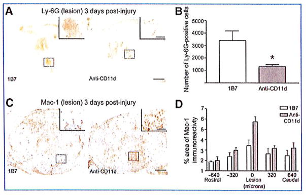
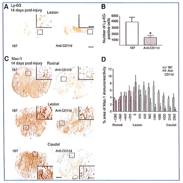
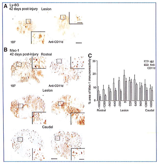
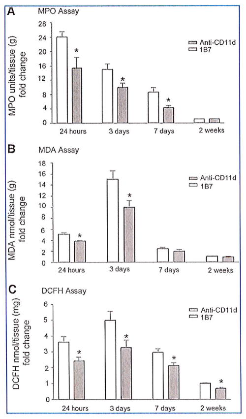
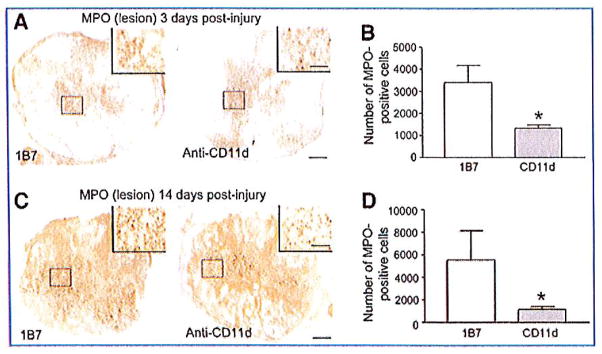
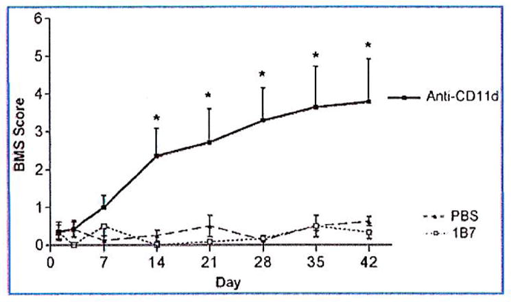
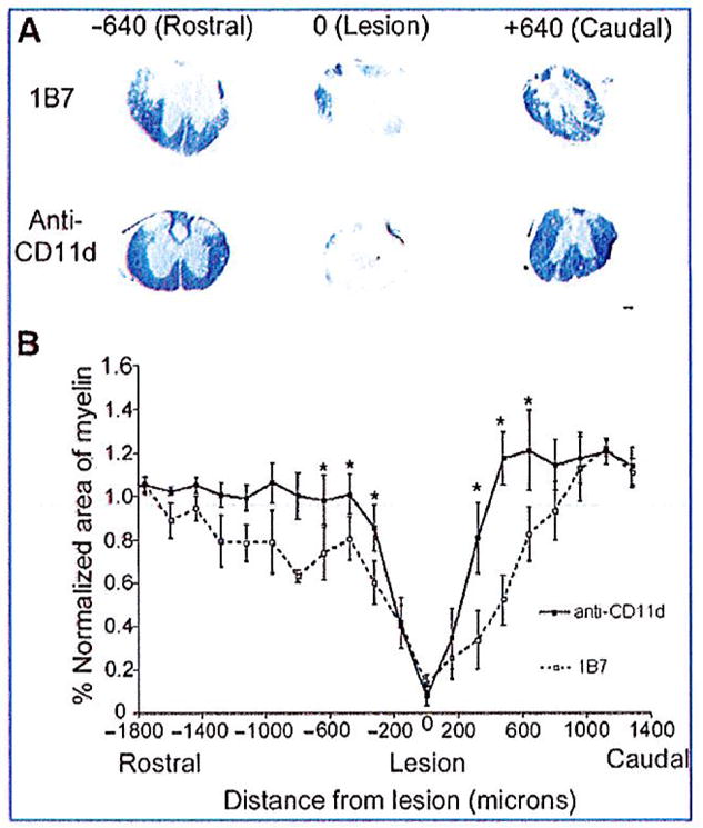
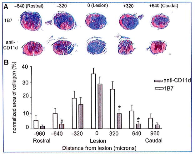
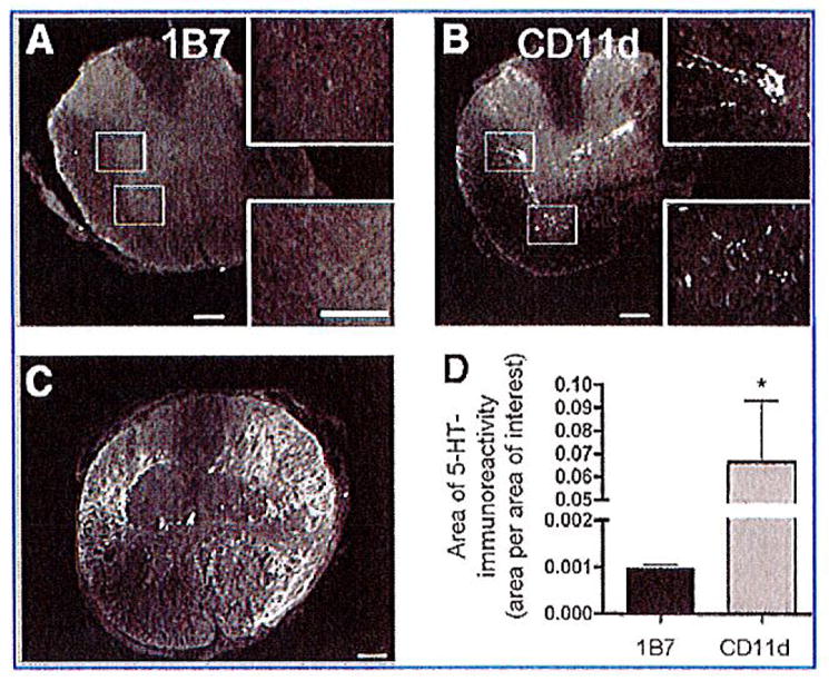
Acute administration of a monoclonal antibody (mAb) raised against the CD11d subunit of the leukocyte CD11d/CD18 integrin after spinal cord injury (SCI) in the rat greatly improves neurological outcomes. This has been chiefly attributed to the reduced infiltration of neutrophils into the injured spinal cord in treated rats. More recently, treating spinal cord-injured mice with a Ly-6G neutrophil-depleting antibody was demonstrated to impair neurological recovery. These disparate results could be due to different mechanisms of action utilized by the two antibodies, or due to differences in the inflammatory responses between mouse and rat that are triggered by SCI. To address whether the anti-CD11d treatment would be effective in mice, a CD11d mAb (205C) or a control mAb (1B7) was administered intravenously at 2, 24, and 48 h after an 8-g clip compression injury at the fourth thoracic spinal segment. The anti-CD11d treatment reduced neutrophil infiltration into the injured mouse spinal cord and was associated with increased white matter sparing and reductions in myeloperoxidase (MPO) activity, reactive oxygen species, lipid peroxidation, and scar formation. These improvements in the injured spinal cord microenvironment were accompanied by increased serotonin (5-HT) immunoreactivity below the level of the lesion and improved locomotor recovery. Our results with the 205C CD11d mAb treatment complement previous work using this anti-integrin treatment in a rat model of SCI.


Conflict of interest statement

Author Disclosure Statement

No competing financial interests exist.

Figures

FIG. 1
FIG. 1
The anti-CD11d treatment reduces neutrophil infiltration into the cord at 3 days post-injury. Shown are representative photomicrographs of sections from the lesion epicenters of 1B7 and CD11d monoclonal antibody (mAb)-treated mice 3 days after spinal cord injury (SCI), immunostained with a Ly-6G antibody (A) or a Mac-1 antibody (C). Staining for neutrophils (Ly-6G) was decreased in mice treated with the CD11d mAb compared to mice treated with the control mAb (1B7). (B) The number of Ly-6G-positive cells at the lesion epicenter in CD11d mAb-treated mice (gray bars) was significantly reduced compared to control 1B7-treated mice (open bars). (D) The mean percent area of Mac-1 immunoreactivity at the lesion epicenter in CD11d mAb-treated mice (gray bars) tended to be increased compared to control 1B7-treated mice (open bars; scale bars in A and C = 100 μm, in high-power inset = 50 μm). In B and D values are means ± standard error (*p<0.05 in B significantly different from controls by two-tailed Student’s t-test, 6 animals/group; p=0.093 in D by two-way analysis of variance, 5 animals/group).
FIG. 2
FIG. 2
CD11d monoclonal antibody (mAb) treatment reduces neutrophil and macrophage/microglia numbers in the injured mouse spinal cord 14 days post-injury. (A and C) Shown are representative photomicrographs of sections from the lesion epicenters of 1B7 and CD11d mAb-treated mice 14 days after spinal cord injury (SCI), immunostained with a Ly-6G antibody. (B) CD11d mAb treatment (gray bar) significantly reduced the number of neutrophils (Ly-6G+ cells) compared to controls (1B7, open bar). (C) Representative photomicrographs of sections at the epicenter or 960 μm caudal or rostral to the epicenter from anti-CD11d or control 1B7 mAb-treated mice stained with a Mac-1 antibody. Mac-1-expressing cells were large, foamy, and round at the lesion epicenters in both CD11d and 1B7 mAb-treated mice, and caudal to the epicenter in 1B7 mAb-treated mice. Rostral to the lesions in both CD11d and 1B7 mAb-treated mice, and caudal to the lesion epicenter in CD11d mAb-treated mice, the majority of Mac-1+ cells were stratified, with small cell bodies bearing multiple processes. (D) CD11d mAb (gray bars) treatment significantly reduced the percent area of Mac-1-immunoreactivity caudal to the lesion compared to controls (1B7, open bars; scale bars in A and C = 100 μm, 50 μm in high-power insets). In B and D values are means ± standard error (in B *p<0.05 significantly different from controls by two-tailed Student’s t-test, 6 mice/group; in D *p=0.025 by two-way analysis of variance and p<0.05 by Fisher protected t-test, 6 mice/group).
FIG. 3
FIG. 3
Macrophage/microglia and neutrophils are seen in the injured cord at 42 days post-injury. (A) Representative photomicrographs of sections from the lesion epicenters of 1B7 and CD11d monoclonal antibody (mAb)-treated mice 42 days after spinal cord injury (SCI), immunostained with a Ly-6G antibody. There was no significant difference in the number of Ly-6G+ cells in CD11d mAb and 1B7 mAb-treated mice 42 days after SCI. (B) Representative photomicrographs of sections at the epicenter or 960 μm caudal or rostral to the epicenter from 1B7 and CD11d mAb-treated mice 42 days after SCI, immunostained with a Mac-1 antibody. Mac-1+ cells were generally stratified in appearance caudal and rostral to the lesion site. At the lesion site, Mac-1 cells were more round in appearance. (C) There was no significant difference in Mac-1 areas of immunoreactivity between 1B7 mAb-treated mice (open bars) compared to anti-CD11d-treated mice (gray bars; 6 mice/1B7 group and 6 mice/anti-CD11d group, two-way analysis of variance; scale bars in A and B = 100 μm and 50 μm for high-power insets).
FIG. 4
FIG. 4
CD11d monoclonal antibody (mAb) treatment decreases inflammation and oxidative damage in the injured spinal cord. (A) Myeloperoxidase (MPO) activity was measured in cord homogenates. CD11d mAb (gray bars) treatment significantly reduced MPO activity at 24 h, 3 days, and 7 days post-injury compared to controls (1B7, open bars). (B) Malondialdehyde (MDA), a measure of lipid peroxidation, was significantly reduced in cord homogenates by the anti-CD11d treatment (gray bars) compared to control treatment (1B7, open bars) at 24 h and 3 days post-injury. (C) 2′-7′-Dichlorofluorescin diacetate (DCFH-DA) oxidation, a measure of reactive oxygen species in injured cord, was significantly reduced by CD11d mAb treatment (gray bars) compared to control treatment (1B7, open bars). Values are means ± standard error (*p<0.05 significantly different from controls by Student’s t-test; n=4 at 24 h; n=6 at 3 days; n=4 at 7 days; n=6 at 2 weeks).
FIG. 5
FIG. 5
Myeloperoxidase (MPO) immunoreactivity in anti-CD11d and 1B7 monoclonal antibody (mAb)-treated mice. MPO-expressing cells were identified by immunostaining at 3 days post-injury (A) and 14 days post-injury (B) at the lesion epicenter. There was a significant decrease in MPO-expressing cells at 3 days post-injury and at 14 days post-injury in CD11d mAb-treated mice compared to 1B7 controls (scale bars = 100 μm and 50 μm for high-power inset). Values are means±standard error; *p<0.05 significantly different from controls by Student’s t-test, 4 animals/group).
FIG. 6
FIG. 6
Locomotor recovery after a spinal cord compression injury. Locomotor function was assessed using the Basso Mouse Scale (BMS). CD11d monoclonal antibody (mAb; black boxes) treatment improved recovery beginning at 14 days post-injury compared to 1B7 controls (open boxes) or saline controls (black triangles). Values are means ± standard error; *significantly different from controls, p< 0.0001 by two-way analysis of variance, and p<0.05 by Fisher’s protected t-test; 6 mice/1B7 group, 4 mice/saline group, 7 mice/anti-CD11d group).
FIG. 7
FIG. 7
CD11d monoclonal antibody (mAb) treatment increases myelin sparing at 42 days after spinal cord injury (SCI). (A) Photomicrographs of solochrome-stained spinal cord sections from 1B7 and CD11d mAb-treated mice rostral (− 640 μm), caudal (+ 640 μm), and at the lesion site (0 μm). Very little myelin (indicated by blue staining) was present at the lesion epicenters. However, myelin increased rostral and caudal to the lesion in CD11d mAb-treated mice. (B) Solochrome staining of consecutive transverse sections from anti-CD11d and 1B7 mAb-treated mice was used to plot the amount of myelin from 1400 μm rostral to 1400 μm caudal to the lesion epicenters. The area of myelin in each section was normalized to the area of myelin in a section with normal morphology from the same cord far from the injury site. CD11d mAb treatment (solid line) increased the amount of myelin beginning at 360 μm rostral and caudal to the lesion compared to control treatment (1B7, dashed line; scale bar in A = 100 μm). Values are means ± standard error (*significantly different from controls, p= 0.008 by two-way analysis of variance, and p<0.05 by Fisher’s protected t-test; 5 mice/1B7 group and 6 mice/anti-CD11d group).
FIG. 8
FIG. 8
CD11d monoclonal antibody (mAb) treatment reduces the collagenous scar 42 days after spinal cord injury. (A) Trichrome staining was used to delineate the presence of collagen (blue color). Representative photomicrographs demonstrate large amounts of collagen at the injury site in CD11d and 1B7 mAb-treated mice. Rostral and caudal to their lesion epicenters, the CD11d mAb-treated mice had reduced levels of blue-stained collagen compared to the 1B7 control section, and almost no collagen was present in sections 640 μm rostral and 640 μm caudal to their lesion epicenters. (B) The area of collagen was reduced with CD11d mAb treatment (gray bars), rostral to the lesion site (−640 μm) and caudal to the lesion site (+320 μm), compared to controls (1B7, open bars; scale bar in A = 100 μm). (B) Values are means ± standard error (*significantly different from controls, p<0.0001 by one-way analysis of variance, and p<0.05 by Student Newman-Keuls test; 5 mice/1B7 group, 6 mice/anti-CD11d group).
FIG. 9
FIG. 9
CD11d monoclonal antibody (mAb) treatment increases serotonin (5-HT) immunoreactivity caudal to the lesion. Immunohistochemistry was used to detect 5-HT in the spinal cord 42 days post-injury. Representative photomicrographs of sections stained for 5-HT immunoreactivity 1920 μm caudal to the lesion epicenter from 1B7- (A) or CD11d-treated (B) mice. (A) Almost no observable 5-HT staining was detected caudal to the lesion in 1B7-treated mice (5 mice/group). (B) Serotonin immunoreactivity was observed caudal to the lesion in the intermediolateral cell column and ventral horn in the injured spinal cords from animals treated with the CD11d mAb (6 mice/group). (C) Representative photomicrograph of a section from the lesion epicenter of a CD11d-treated mouse stained for 5-HT immunoreactivity. The appearance of the 5-HT immunoreactivity in the lesion epicenters of 1B7 mAb-treated controls was no different from the appearance of the 5-HT immunoreactivity seen in the CD11d mAb-treated mice (scale bars = 100 μm). (D) Measuring the area of 5-HT-immunoreactivity (area/area of interest) at 1920 μm caudal to the lesion epicenter in CD11d- and 1B7-treated spinal cord-injured mice revealed a significant increase in 5-HT immunoreactivity in the CD11d mAb-treated group.

References

    1. Bao F, Chen Y, Dekaban GA, Weaver LC. An anti-CD11d integrin antibody reduces cyclooxygenase-2 expression and protein and DNA oxidation after spinal cord injury in rats. J Neurochem. 2004;90:1194–1204. - PubMed
    1. Bao F, Chen Y, Dekaban GA, Weaver LC. Early anti-inflammatory treatment reduces lipid peroxidation and protein nitration after spinal cord injury in rats. J Neurochem. 2004;88:1335–1344. - PubMed
    1. Bao F, Dekaban GA, Weaver LC. Anti-CD11d antibody treatment reduces free radical formation and cell death in the injured spinal cord of rats. J Neurochem. 2005;94:1361–1373. - PubMed
    1. Bartholdi D, Schwab ME. Methylprednisolone inhibits early inflammatory processes but not ischemic cell death after experimental spinal cord lesion in the rat. Brain Res. 1995;672:177–186. - PubMed
    1. Basso DM, Fisher LC, Anderson AJ, Jakeman LB, McTigue DM, Popovich PG. Basso Mouse Scale for locomotion detects differences in recovery after spinal cord injury in five common mouse strains. J Neurotrauma. 2006;23:635–659. - PubMed

This article has been cited by

    1. Zhang B, Gensel JC. Is neuroinflammation in the injured spinal cord different than in the brain? Examining intrinsic differences between the brain and spinal cord. Experimental Neurology. 2014;258:112–120. - PubMed
    1. Bastien Dominic, Lacroix Steve. Cytokine pathways regulating glial and leukocyte function after spinal cord and peripheral nerve injury. Experimental Neurology. 2014;258:62–77. - PubMed
    1. Nardone Raffaele, Höller Yvonne, Thomschewski Aljoscha, Höller Peter, Lochner Piergiorgio, Golaszewski Stefan, Brigo Francesco, Trinka Eugen. Serotonergic transmission after spinal cord injury. Journal of Neural Transmission 2014 - PubMed
    1. John C. GenselSpinal Cord Injury. pp. 339–361.
    1. Vaughn Chloe N, Iafrate Julia L, Henley Jessica B, Stevenson Edward K, Shlifer Igor G, Bucky Jones T. Cellular Neuroinflammation in a Lateral Forceps Compression Model of Spinal Cord Injury. The Anatomical Record. 2013;296(8):1229–1246. - PubMed

Publication types

Grants and funding