. 2012 Jan-Feb;88(1):119-28.
doi: 10.1111/j.1751-1097.2011.01027.x. Epub 2011 Nov 29.

Diversity of Chlamydomonas channelrhodopsins

Affiliations

Diversity of Chlamydomonas channelrhodopsins

Sing-Yi Hou et al. Photochem Photobiol. 2012 Jan-Feb.

Abstract

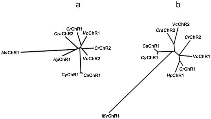
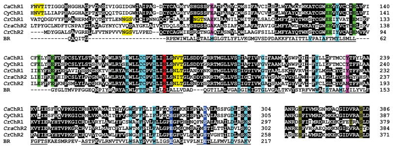
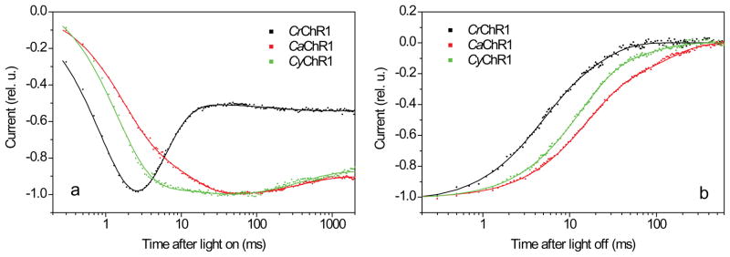
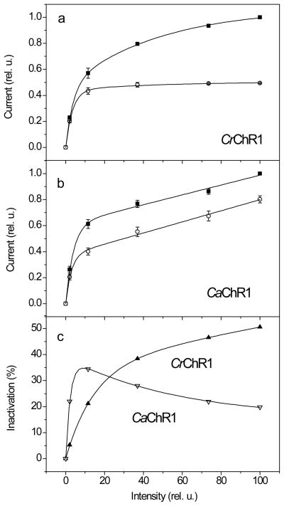
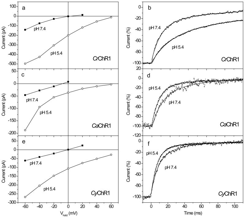
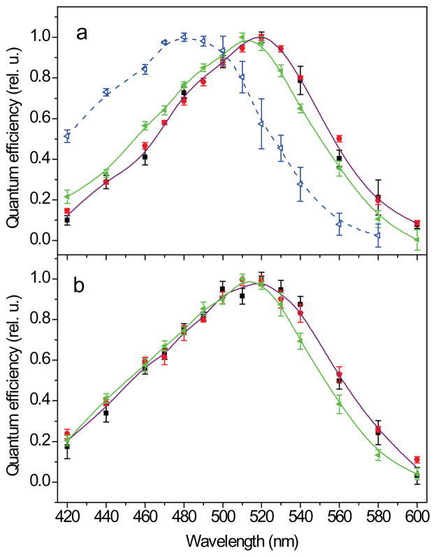
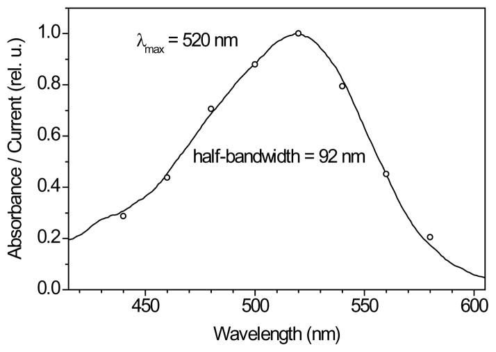
Channelrhodopsins act as photoreceptors for control of motility behavior in flagellates and are widely used as genetically targeted tools to optically manipulate the membrane potential of specific cell populations ("optogenetics"). The first two channelrhodopsins were obtained from the model organism Chlamydomonas reinhardtii (CrChR1 and CrChR2). By homology cloning we identified three new channelrhodopsin sequences from the same genus, CaChR1, CyChR1 and CraChR2, from C. augustae, C. yellowstonensis and C. raudensis, respectively. CaChR1 and CyChR1 were functionally expressed in HEK293 cells, where they acted as light-gated ion channels similar to CrChR1. However, both, which are similar to each other, differed from CrChR1 in current kinetics, inactivation, light intensity dependence, spectral sensitivity and dependence on the external pH. These results show that extensive channelrhodopsin diversity exists even within the same genus, Chlamydomonas. The maximal spectral sensitivity of CaChR1 was at 520 nm at pH 7.4, about 40 nm redshifted as compared to that of CrChR1 under the same conditions. CaChR1 was successfully expressed in Pichia pastoris and exhibited an absorption spectrum identical to the action spectrum of CaChR1-generated photocurrents. The redshifted spectra and the lack of fast inactivation in CaChR1- and CyChR1-generated currents are features desirable for optogenetics applications.


Figures

Figure 1
Figure 1
Phylogenetic trees of the 7TM domains (a) and the C-terminal domains (b) of the so far known channelopsins constructed by the neighbor-joining method. CrChR1, channelrhodopsin 1 from C. reinhardtii; CrChR2, channelrhodopsin 2 from C. reinhardtii; VcChR1, channelrhodopsin 1 from V. carteri; VcChR2, channelrhodopsin 2 from V. carteri; MvChR1, channelrhodopsin 1 from M. viride; CaChR1, channelrhodopsin 1 from C. augustae; CyChR1, channelrhodopsin 1 from C. yellowstonensis; CraChR2, channelrhodopsin 2 from C. raudensis; HpChR1 channelrhodopsin 1 from Haematococcus pluvialis.
Figure 2
Figure 2
Partial alignment of Chlamydomonas channelopsin and BR sequences. Black background indicates conserved identical residues. Turquoise background, positions of the residues that form the retinal-binding pocket in BR. Green background, conserved Glu residues in the predicted second helix. Magenta background, molecular determinants that differentiate CrChR1/VcChR1 from CrChR2/VcChR2. Red background, residues in the position of the proton donor in BR. Blue background, residues in the positions of Glu194 and Glu204 in BR. Yellow background, predicted glycosylation sites. Olive background, conserved residues known to be phosphorylated in CrChR1 or CrChR2. Underlined characters show the regions that form transmembrane helices in BR.
Figure 3
Figure 3
(a) Typical kinetics of light-induced currents generated in HEK293 cells by CrChR1 (black dots), CaChR1 (red dots) and CyChR1 (green dots). (b) Decay of the same currents after 2-s illumination. The currents in (a) were normalized to the peak amplitude, and the currents in (b), to the plateau level, and fitted with three (a) or two (b) exponential functions (solid lines). The excitation wavelength was 520 nm for CaChR1 and CyChR1, and 480 nm for CrChR1, which corresponded to their spectral maxima (see below). The traces are average signals measured in response to a series of light pulses delivered with 30-s time intervals. Cells expressing ChRs were selected for EYFP fluorescence before measurements. Bath pH was 7.4, Vhold was -60 mV. For complete ionic conditions, see Methods section.
Figure 4
Figure 4
(a, b) The dependence of peak (solid squares) and plateau (open circles) amplitudes on the stimulus intensity for currents generated by CrChR1 (a) or CaChR1 (b). Data points are the mean normalized values ± SEM (n = 3 (a) and 5 (b)). For complete ionic conditions, see Methods section. (c) The dependence of light inactivation (calculated as the difference between the peak and plateau amplitudes shown in panels (a) and (b), relative to the peak amplitude) on the stimulus intensity for currents generated by CrChR1 (solid triangles) and CaChR1 (open triangles).
Figure 5
Figure 5
(a, c, e) Typical current-voltage relationships (I-V curves) for the plateau level measured at the end of a 2-s excitation light pulse upon an increase of Vhold in 20 mV steps from −60 mV at the bath pH 7.4 (solid squares) and 5.4 (open circles) in HEK293 cells transfected with CrChR1, CaChR1, or CyChR1. The wavelength was 520 nm for CaChR1 and CyChR1, and 480 nm for CrChR1, which corresponded to their spectral maxima (see below). (b, d, e) Normalized current decay traces recorded from cells transfected with CrChR1, CaChR1, or CyChR1 at holding potential (Vhold) −60 mV. Traces at the bath pH 7.4 or 5.4 (indicated in the panels) were recorded from the same cell. Note the opposite effects of pH changes on the decay kinetics in CrChR1 and the new channelrhodopsins. Zero time corresponds to the end of a 2-s excitation light pulse. Excitation light was as in a, c, e. Experimental data (dots) were fitted with two exponential functions (solid lines).
Figure 6
Figure 6
(a, b) The action spectra of photoelectric currents generated in HEK293 cells by CaChR1 (a) or CyChR1 (b) at the bath pH 7.4 (black squares), 5.4 (red circles) or 9.0 (green solid triangles). For comparison, the action spectrum of ChR1 from C. reinhardtii measured at pH 7.4 is shown in panel A (blue open triangles, dashed line).
Figure 7
Figure 7
Comparison of the absorption spectrum of partially purified CaChR1 in detergent (black line) with the action spectrum of photocurrents (open circles), pH 7.4.

References

    1. Bogomolni R, Spudich JL. Identification of a third rhodopsin-like pigment in phototactic Halobacterium halobium. Proc Natl Acad Sci USA. 1982;79:6250–6254. - PMC - PubMed
    1. Spudich JL, Bogomolni RA. Mechanism of colour discrimination by a bacterial sensory rhodopsin. Nature. 1984;312:509–513. - PMC - PubMed
    1. Foster KW, Saranak J, Patel N, Zarrilli G, Okabe M, Kline T, Nakanishi K. A rhodopsin is the functional photoreceptor for phototaxis in the unicelullar eukaryote Chlamydomonas. Nature. 1984;311:756–759. - PubMed
    1. Asamizu E, Miura K, Kucho K, Inoue Y, Fukuzawa H, Ohyama K, Nakamura Y, Tabata S. Generation of expressed sequence tags from low-CO2 and high-CO2 adapted cells of Chlamydomonas reinhardtii. DNA Research. 2000;7:305–307. - PubMed
    1. Sineshchekov OA, Jung KH, Spudich JL. Two rhodopsins mediate phototaxis to low- and high-intensity light in Chlamydomonas reinhardtii. Proc Natl Acad Sci USA. 2002;99:8689–8694. - PMC - PubMed

Publication types

LinkOut - more resources