. 2012 Jan 5;422(1):59-69.
doi: 10.1016/j.virol.2011.09.033. Epub 2011 Oct 30.

Molecular characterization of the newly identified human parvovirus 4 in the family Parvoviridae

Affiliations

Molecular characterization of the newly identified human parvovirus 4 in the family Parvoviridae

Sai Lou et al. Virology. .

Abstract

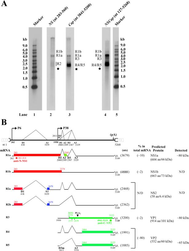
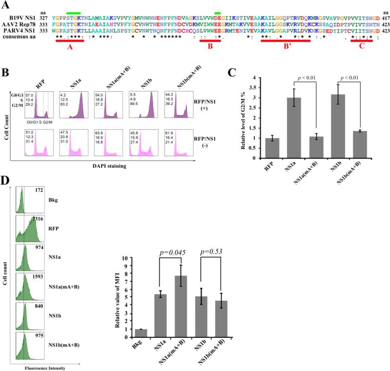
Human parvovirus 4 (PARV4) is an emerging human virus, and little is known about the molecular aspects of PARV4 apart from its incomplete genome sequence, which lacks information of the termini. We analyzed the gene expression profile of PARV4 using a nearly full-length HPV4 genome in a replication competent system in 293 cells. We found that PARV4 utilizes two promoters to transcribe non-structural protein- and structural protein-encoding mRNAs, respectively, which were polyadenylated at the right end of the genome. Three major proteins, including the large non-structural protein NS1a, whose mRNA is spliced, and capsid proteins VP1 and VP2, were detected. Additional functional analysis of the NS1a revealed its capability to induce cell cycle arrest at G2/M phase in ex vivo-generated human hematopoietic stem cells. Taken together, our characterization of the molecular features of PARV4 suggests that PARV4 represents a new genus in the family Parvoviridae.


Figures

Figure 1
Figure 1. Replication of the construct p5TRPARV4 in 293 cells
293 cells were transfected with p5TRPARV4, pAV5Rep78 and pHelper, or pSKPARV4 alone. Hirt DNA was extracted from the cells 48 hrs post-transfection and digested with DpnI (DpnI +) or without DpnI (DpnI -). The DNA samples were then subjected to Southern blot analysis using the PARV4 NSCap probe (nt 127-5268). Ten ng of the probe template DNA was treated in parallel as a DpnI digestion control (lanes 1& 2).
Figure 2
Figure 2. 5’/3’ RACE and RT-PCR analyses of PARV4 mRNAs
Total RNA was isolated from 293 cells co-transfected with p5TRPARV4, pAV5Rep78, and pHelper and subjected to reverse transcription. The cDNA was then subjected to 5’/3’ RACE analysis or amplified with PARV4-specific primers. (A) Amplified DNA fragments were electrophoresed on 2% agarose gel and visualized using ethidium bromide staining. (B) Primer sets used for PCR amplification are depicted, and the transcription units identified, with the promoters (P6 and P38), splice donor (D1, D2, D3 and D3m) and acceptor (A1, A2 and A3) sites, and polyadenylation signals [(pA)p and (pA)d ], are shown. LTR, left terminal repeat; RTR, right terminal repeat.
Figure 3
Figure 3. Transcription mapping by RNase protection assays (RPAs)
(A) Schematic diagram of the PARV4 genome and the probes used for the RPA. The PARV4 genome is depicted with the transcription units. Probes used for the RPAs are shown with their respective nucleotide numbers, together with the bands that they are expected to protect and the predicted sizes. Spl, spliced mRNAs; Unspl, unspliced mRNAs; RT, mRNAs read through the polyadenylation site. (B) Mapping of the PARV4 transcripts by RPA. Ten μg of total RNA was isolated from 293 cells transfected with p5TRPARV4, pAV5Rep78, and pHelper, and were protected by PARV4 probes P1 and P3-9. Lanes 1&9, 32P-labeled RNA markers with sizes indicated. Bands detected are designated in each lane.
Figure 4
Figure 4. Transcription profile of PARV4
(A) Northern blot analysis. Total RNA was isolated from 293 cells transfected with p5TRPARV4, pAV5Rep78, and pHelper, and used for Northern blot analysis. The blots were hybridized to three PARV4 DNA probes (NSCap, NS, and Cap), respectively. Bands of mRNA detected by each probe are designated in each lane. The RNA marker ladder is shown in lanes 1 and 5. The asterisk denotes a likely non-specific band detected by all three probes. (B) Transcription map of PARV4. The PARV4 genome is shown to scale, with transcription landmarks indicated. Major mRNA species detected in RT-PCR, RPA, and Northern blot assays are diagrammed to display their identities and respective sizes in the absence of the polyA tail. The putative ORFs that each can encode are also diagramed in boxes, and the predicted sizes (kDa) of translated proteins are indicated on the right. The relative abundance of each mRNA and proteins detected by transfection are also shown on the right. Potential AUG usage for VP1 translation and peptides used for production of antibodies are depicted. N/D, not determined or not detected.
Figure 5
Figure 5. Western blot analysis of PARV4 proteins
293 cells were transfected with plasmids as indicated. At 48 hrs post-transfection, the cells were harvested and lysed for Western blotting using polyclonal antibodies anti-N-NS1 (A) and anti-C-VP2 (B), respectively. The blots were reprobed using an anti-β-actin antibody. The identities of detected proteins are shown to the right on the blot. Untransfected cells were used as a control. The asterisk indicates bands that were likely degraded from VP2.
Figure 6
Figure 6. The helicase motifs of the PARV4 NS1 are responsible for G2/M arrest induced by PARV4 NS1 protein
(A) Comparison of the NS1 helicase motifs. The amino acid sequences of B19V NS1, AAV2 Rep78, and PARV4 NS1 were aligned using ClustalW2 (Larkin et al., 2007). The alignment of aa 327-417 of B19V NS1, aa 333-423 of AAV2 Rep78, and aa 333-423 of PARV4 NS1 is depicted with identical amino acids shown by the asterisk, while homologous amino acids are shown as two dots under the amino acid. Conserved motifs (Walker boxes A, B, B’, and C) are underlined in red, while the mutated regions of boxes A and B are marked in green. (B&C) PARV4 NS1 protein induces G2/M arrest. (B) CD34+ HSCs were transduced with lentiviruses to express proteins as indicted. At 48 hrs post-transduction, RFP- or NS1-expressing cells were selectively gated for anti-Flag staining, followed by DAPI staining for cell cycle analysis. The cell cycle patterns of both NS1/RFP-positive and NS1/RFP-negative cells are shown in parallel. In each panel, the percentages of cells in the G1, S, and G2/M phases are indicated, respectively (top left). A representative experiment is shown. (C) The percentage of RFP-expressing cells in G2/M was arbitrarily set as 1. Relative values are shown with average and standard deviation from at least three independent experiments. P values were determined using a student's t test. (D) Expression levels of PARV4 NS1 proteins. The expression levels of RFP and NS1 as mean fluorescence intensity (MFI) were analyzed by flow cytometry in lentivirus-transduced (NS1/RFP+) cells at 48 hrs post-transduction. The reference line is selected arbitrarily to show the relative position of the NS1/RFP-positive and negative peaks, and Bkg (background) represents the secondary antibody only control. A representative experiment is shown to the left. Relative MFI values were determined as fold changes of MFI of NS1/RFP+ cells compared to that of Bkg, and are shown to the right with average and standard deviation.
Figure 7
Figure 7. The PARV4 VP1 unique region does not exhibit phospholipase A2-like activity in vitro
(A) Comparison of B19V VP1u and PARV4 VP1u. The PARV4 VP1 and potential unique regions are diagramed. The alignment of PLA2-conserved motifs of B19V VP1u and three forms of PARV4 VP1u is depicted, with identical amino acids shown as an asterisk and homologous amino acids shown as two dots. (B) SDS-PAGE analysis of purified PARV4-VP1u and B19V VP1u proteins. Five microliter of each purified protein and BSA standards was analyzed on SDS-10%PAGE gel. The gel was stained using Coomassie blue. A protein maker is shown. (C) PARV4 VP1u does not exhibit PLA2-like activity. An in vitro sPLA2 assay kit was used to test PLA2-like activity of various substrates as indicated on the right. The PLA2-like activity is shown as a value of the absorbance at 414 nm at various time points. BLK, blank.

References

    1. Adlhoch C, Kaiser M, Ellerbrok H, Pauli G. High prevalence of porcine Hokovirus in German wild boar populations. Virol. J. 2010;7:171. 171. - PMC - PubMed
    1. Ahn JK, Pitluk ZW, Ward DC. The GC box and TATA transcription control elements in the P38 promoter of the minute virus of mice are necessary and sufficient for transactivation by the nonstructural protein NS1. J. Virol. 1992;66:3776–3783. - PMC - PubMed
    1. Allander T, Tammi MT, Eriksson M, Bjerkner A, Tiveljung-Lindell A, Andersson B. Cloning of a human parvovirus by molecular screening of respiratory tract samples. Proc. Natl. Acad. Sci. U. S. A. 2005;102:12891–12896. - PMC - PubMed
    1. Anderson S, Momoeda M, Kawase M, Kajigaya S, Young NS. Peptides derived from the unique region of B19 parvovirus minor capsid protein elicit neutralizing antibodies in rabbits. Virology. 1995;206:626–632. - PubMed
    1. Benjamin LA, Lewthwaite P, Vasanthapuram R, Zhao G, Sharp C, Simmonds P, Wang D, Solomon T. Human parvovirus 4 as potential cause of encephalitis in children, India. Emerg. Infect. Dis. 2011;17:1484–1487. - PMC - PubMed

Publication types