Review
. 2012 Aug 15;62(2):620-31.
doi: 10.1016/j.neuroimage.2011.10.044. Epub 2011 Oct 20.

Sewer pipe, wire, epoxy, and finger tapping: the start of fMRI at the Medical College of Wisconsin

Affiliations
Review

Sewer pipe, wire, epoxy, and finger tapping: the start of fMRI at the Medical College of Wisconsin

Peter A Bandettini. Neuroimage. .

Abstract

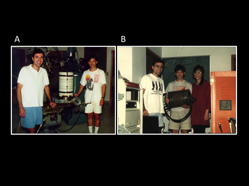
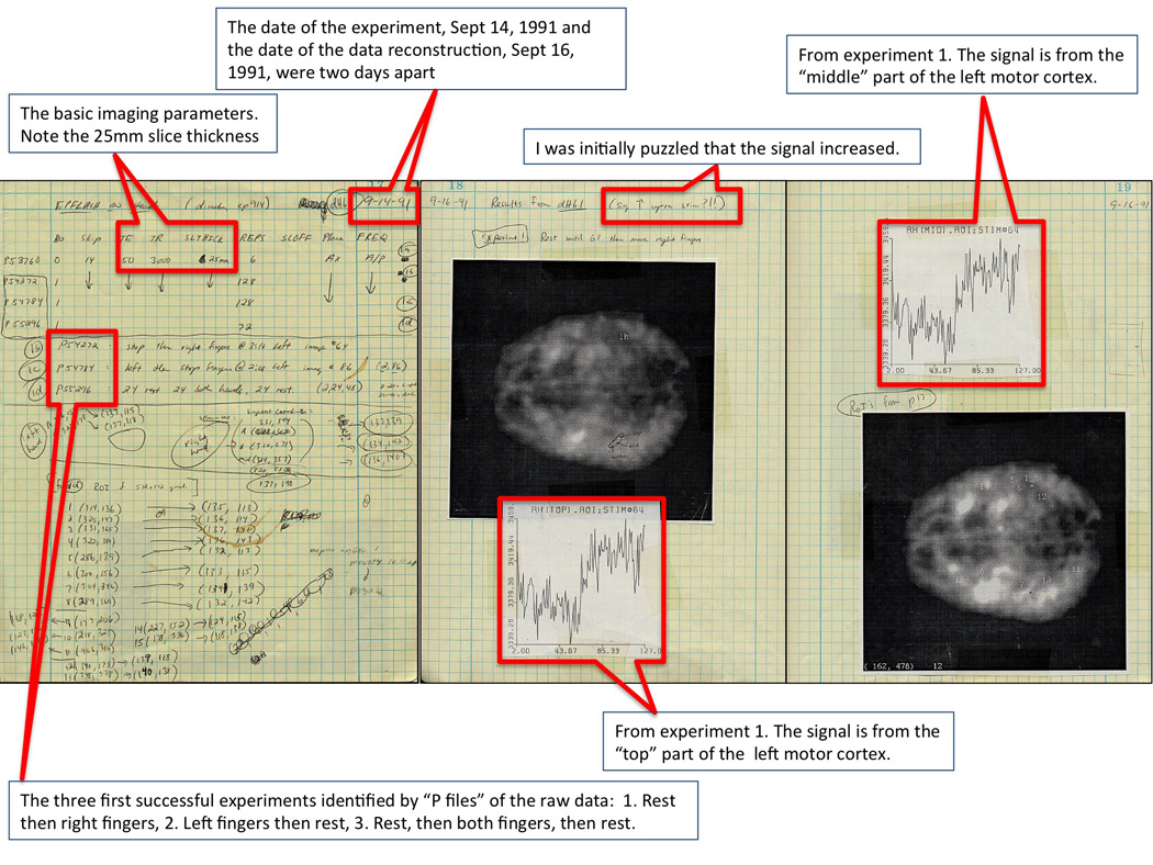
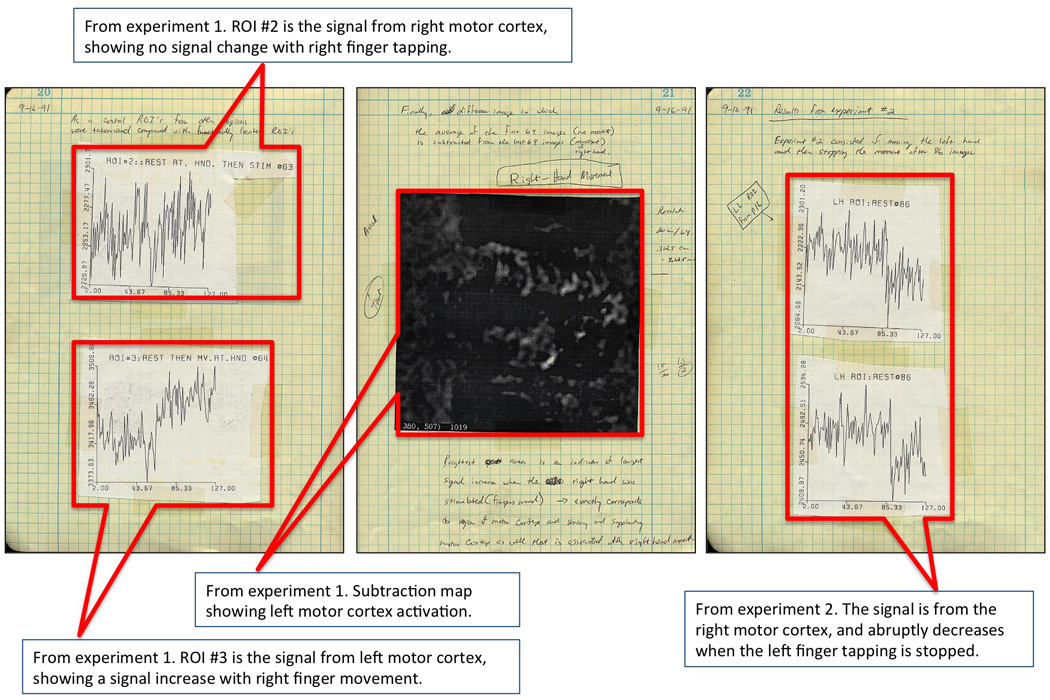
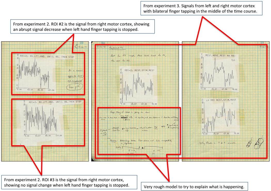
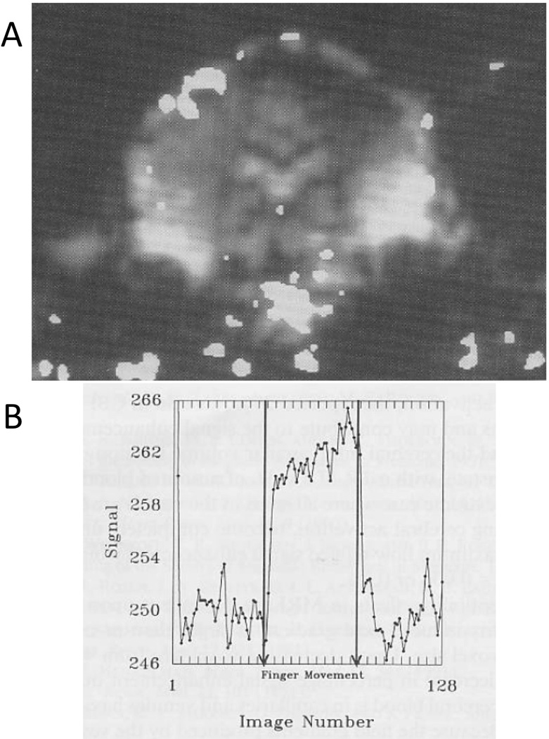
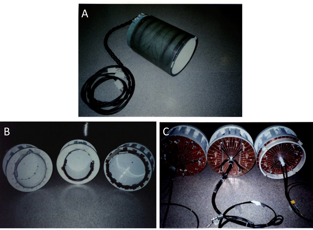
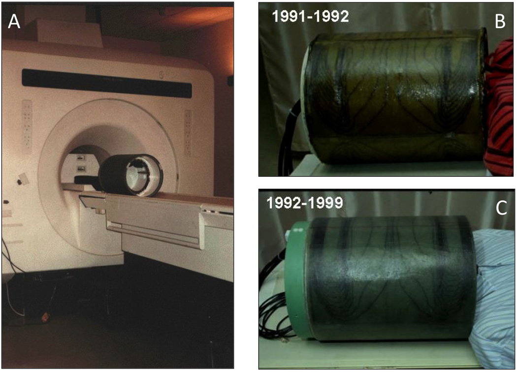
In 1991, the Biophysics Research Institute at the Medical College of Wisconsin was among the first groups to develop functional Magnetic Resonance Imaging (fMRI). Our story is unique on a few levels: We didn't have knowledge of the ability to image human brain activation with MRI using blood oxygenation dependent (BOLD) contrast until early August of 1991 when we attended the Society for Magnetic Resonance in Medicine (SMRM) meeting in San Francisco, yet we produced our first BOLD-based maps of motor cortex activation about a month later. The effort started with two graduate students, Eric Wong and myself. Only a few days prior to that extremely important SMRM meeting, we had developed human echo planar imaging (EPI) capability in-house. Wong designed, built, and interfaced a head gradient coil made out of sewer pipe, wire, and epoxy to a standard GE 1.5T MRI scanner. Also, a few months prior to building this human head gradient coil he developed the EPI pulse sequences and image reconstruction. All of these efforts were towards a different goal--for demonstration of Wong's novel approach to perfusion imaging in the human brain. Following SMRM, where a plenary lecture by Tom Brady from MGH opened our eyes to human brain activation imaging using BOLD contrast, and where we learned that EPI was extremely helpful if not critical to its success, we worked quickly to achieve our first results on September 14, 1991. The story is also unique in that Jim Hyde had set up the Biophysics Research Institute to be optimal for just this type of rapidly advancing basic technology research. It was well equipped for hardware development, had open and dynamic collaborative relationships with other departments, hospitals on campus, and GE, and had a relatively flat hierarchy and relaxed, flexible, collegial atmosphere internally. Since these first brain activation results, MCW Biophysics has continued to be at the forefront of functional MRI innovation, having helped to pioneer real time fMRI, high-resolution fMRI, and functional connectivity mapping.


Figures

Figure 1
Figure 1
These were taken during the first week of August, 1991. They show: A. Eric Wong and myself during the early stages of the construction of the three-axis balanced torque human head gradient coil that was used for the first collection of fMRI data at MCW. Here, the coil has the first layer of wires (z-axis) applied. B. Here, Eric, his wife Denise, and I show the coil after collecting the first image (an apple) with it. It was interfaced to a GE 1.5T Signa scanner. This picture was taken after we’ve been working almost continuously for about 36 hours to complete it.
Figure 2
Figure 2
These are the first three of nine pages from my lab notebook, showing our first successful results. The experiments were performed Sept 14, 1991 and the data were finally reconstructed and analyzed Sept 16, 1991. The first three experiments were of a very thick (25 mm) axial slice through the motor cortex of my brain. The signal clearly goes up in the contralateral motor cortex during finger tapping. I was initially puzzled that the signal should increase, but then, on a review of PET and optical imaging literature, giving evidence that with activation, oxygen extraction fraction went down, it all made sense.
Figure 3
Figure 3
These are the second three of nine pages from my lab notebook, showing our first successful results. I show control data showing no signal change from the ipsalateral motor cortex and also show a subtraction map, revealing a signal increase in left motor cortex during right finger tapping.
Figure 4
Figure 4
These are the last three pages of the nine from my lab notebook, showing our first successful results. They include more time series, and, specifically, a time series showing our first true block design: rest, bilateral finger tapping, and then rest again. I also started doodling a rough model for what I thought was going on.
Figure 5
Figure 5
These are the results from the GE prototype z-axis head gradient coil. During this experiment (Alan Song was the volunteer), I had to pull a string attached to his hand to get him to tap his fingers. A. This is a functional map that was “thresholded” or “threshheld” (these really should become words since they are used so much in fMRI) and superimposed on an EPI anatomic image from the time series. Because the gradient coil was z-axis, we had to collect either sagittal or coronal EPI slices. Note the large pulsation artifact at the base of the brain. B. The signal to noise of the GE body RF coil was a better than that of the first generation RF coil inside the gradient coil. The time series signal from a region of interest in the motor cortex looks less contaminated by noise. (Reproduced with permission from Wiley)
Figure 6
Figure 6
A. Our second-generation gradient coil. It was a bit larger and had the option to switch the z-axis windings from parallel to series to double the gradient strength in z for diffusion weighting – at the expense of slew rate. One advantage was that it allowed the insertion of different RF coils. The 0.5T, 1.5T, and 3T coils are shown in B and C. C shows that they were all end-capped with slits in the copper end-caps placed to minimize eddy currents.
Figure 7
Figure 7
A. This is a picture of the balanced-torque gradient coil as it looked when it was about to be used. We only added a patient restraint band when using it. B and C show the two gradient coils being modeled by me – quite comfortable. The years that they were used are shown in the upper left of each picture.
Figure 8
Figure 8
This fMRI-based brain activation movie of alternating left and right finger tapping was first presented at the SMRI meeting in New York in April of 1992. It is an axial slice and consists of a series of images that were subtracted from a baseline image. It is approximately 5 × faster than real time and clearly shows right then left motor cortex becoming active associated with right then left finger tapping.
Figure 9
Figure 9
The picture (from Bob Cox’s current NIH office wall) of the email sent from Bob to Jim Hyde telling him that he was stopping development of AFNI – back in November 1994, and about a year after he started…he had barely just begun.
Figure 10
Figure 10
A picture of the “early adopters” of fMRI at MCW with Jim Hyde, taken at the first Organization for Human Brain Mapping (OHBM) meeting in Paris in June, 1995. From left are Ted DeYoe (focus was vision), Elliot Stein (focus was drug effects), Jim Hyde, Steve Rao (focus was motor activation), and Jeff Binder (focus was language and auditory processing).

References

    1. Bandettini PA, Jesmanowicz A, Van Kylen J, Birn RM, Hyde JS. Functional MRI of brain activation induced by scanner acoustic noise. Magnetic Resonance in Medicine. 1998;39:410–416. - PubMed
    1. Bandettini PA, Jesmanowicz A, Wong EC, Hyde JS. Processing Strategies for Time-Course Data Sets in Functional Mri of the Human Brain. Magnetic Resonance in Medicine. 1993;30:161–173. - PubMed
    1. Bandettini PA, Wong EC. Effects of Biophysical and Physiological- Parameters on Brain Activation-Induced R(2)Asterisk and R(2) Changes - Simulations Using a Deterministic Diffusion-Model. International Journal of Imaging Systems and Technology. 1995;6:133–152.
    1. Bandettini PA, Wong EC. A hypercapnia-based normalization method for improved spatial localization of human brain activation with fMRI. Nmr in Biomedicine. 1997;10:197–203. - PubMed
    1. Bandettini PA, Wong EC, Hinks RS, Tikofsky RS, Hyde JS. Time course EPI of human brain function during task activation. Magnetic resonance in medicine : official journal of the Society of Magnetic Resonance in Medicine / Society of Magnetic Resonance in Medicine. 1992;25:390–397. - PubMed