. 2012 Mar;20(3):625-32.
doi: 10.1038/mt.2011.231. Epub 2011 Nov 1.

Correction of murine Bernard-Soulier syndrome by lentivirus-mediated gene therapy

Affiliations

Correction of murine Bernard-Soulier syndrome by lentivirus-mediated gene therapy

Sachiko Kanaji et al. Mol Ther. 2012 Mar.

Abstract

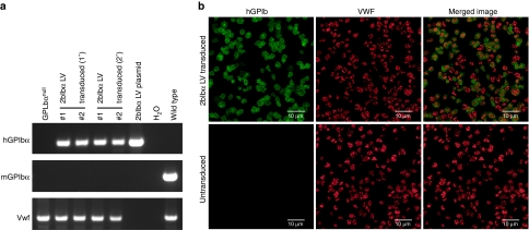
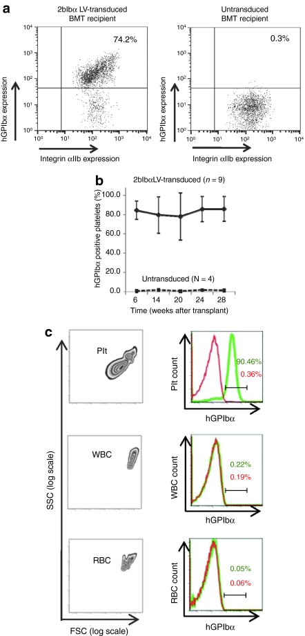
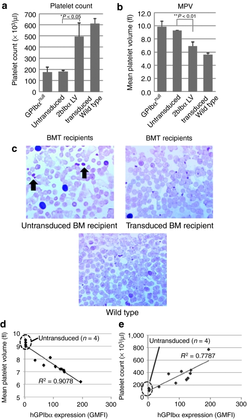
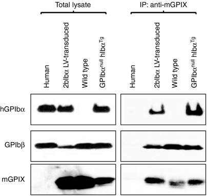
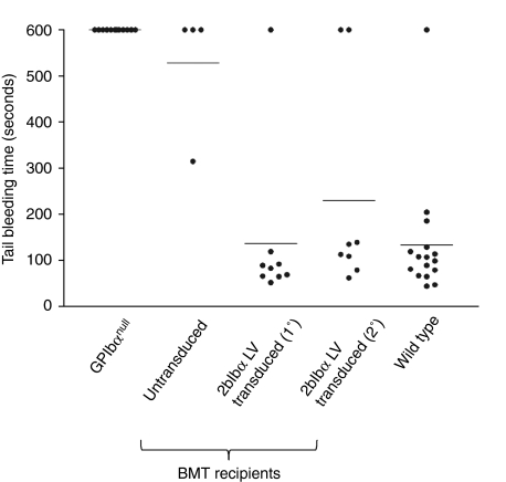
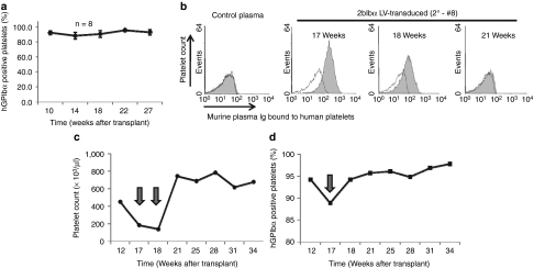
Bernard-Soulier syndrome (BSS) is an inherited bleeding disorder caused by a defect in the platelet glycoprotein (GP) Ib-IX-V complex. The main treatment for BSS is platelet transfusion but it is often limited to severe bleeding episodes or surgical interventions due to the risk of alloimmunization. We have previously reported successful expression of human GPIbα (hGPIbα) in human megakaryocytes using a lentiviral vector (LV) encoding human GP1BA under control of the platelet-specific integrin αIIb promoter (2bIbα). In this study, we examined the efficacy of this strategy for the gene therapy of BSS using GPIbα(null) as a murine model of BSS. GPIbα(null) hematopoietic stem cells (HSC) transduced with 2bIbα LV were transplanted into lethally irradiated GPIbα(null) littermates. Therapeutic levels of hGPIbα expression were achieved that corrected the tail bleeding time and improved the macrothrombocytopenia. Sequential bone marrow (BM) transplants showed sustained expression of hGPIbα with similar phenotypic correction. Antibody response to hGPIbα was documented in 1 of 17 total recipient mice but was tolerated without any further treatment. These results demonstrate that lentivirus-mediated gene transfer can provide sustained phenotypic correction of murine BSS, indicating that this approach may be a promising strategy for gene therapy of BSS patients.


Figures

Figure 1
Figure 1
Genetic and expression analysis of 2bIbαLV-transduced bone marrow transplantation (BMT) recipients. (a) PCR analysis of BMT recipients shows the presence of transgene in recipients. Genomic DNA was prepared from primary (1°) and secondary (2°) 2bIbα lentiviral vector (LV) transduced hematopoietic stem cells (HSC) recipients. GPIbαnull and C57BL/6J wild-type mice were used as controls. 2bIbα LV plasmid DNA was used as a positive control for human GPIbα (hGPIbα). Absence of mGPIbα PCR product confirmed the GPIbαnull background. The Vwf gene was used as an internal control. PCR product sizes; hGPIbα (458 bp), mGPIbα (486 bp), and Vwf (727 bp). (b) Immunofluorescent staining of mouse platelets. Platelets were isolated from GPIbαnull mice that received 2bF8 LV-transduced HSC (upper panel) and untransduced GPIbαnull control mice (lower panel) and stained for hGPIbα (green) and murine VWF (red). Nonspecific isotype-matched primary antibodies were used to assess background staining (data not shown). Bar = 10 µm.
Figure 2
Figure 2
Flow cytometric analysis of human GPIbα (hGPIbα) expression in bone marrow transplantation (BMT) recipients. (a) Expression of hGPIbα in the platelets of 2bIbα lentiviral vector (LV) transduced (upper panel) and untransduced (lower panel) hematopoietic stem cells (HSC) recipients were analyzed by flow cytometry. The platelet population was gated with anti-mouse CD41/integin αIIb mAb and hGPIbα expression was analyzed using AlexaFluor 647 labeled anti-hGPIbα mAb (AP1). (b) Expression of hGPIbα was monitored for 28 weeks after BMT and average expression (percentage of hGPIbα-positive platelets) in 2bIbα LV-transduced HSC recipients (n = 9) was plotted at each time point. Untransduced controls (n = 4) were analyzed in parallel each time. Data is expressed as the mean ± SD. (c) Platelet-specific expression of hGPIbα. Entities exhibiting the forward (FSC) and side (SSC) scattering properties of platelets (Plt), white blood cells (WBC), and red blood cells (RBC) from whole blood of 2bIbα LV-transduced HSC recipients (left columns) were gated to analyze hGPIbα expression on the various blood cell populations. Right columns show histograms of hGPIbα expression in transduced (green) and untransduced (red) HSC recipients. Only platelets from transduced recipient display hGPIbα on their surface.
Figure 3
Figure 3
Analysis of platelet count and size. (a) Platelet count and (b) size of GPIbαnull (n = 6), untransduced bone marrow (BM) recipients (n = 4), 2bIbα lentiviral vector (LV)-transduced BM recipients (n = 9), and C57BL/6J wild-type mice (n = 6) are depicted. Both parameters were monitored every 4 weeks after bone marrow transplantation (BMT) and representative data (20 weeks after BMT) are shown. Data is expressed as the mean ± SD. (c) Blood smears were stained with hematoxylin and eosin. Arrows show giant platelets that were observed in untransduced BMT recipients. (d) Expression of human GPIbα (hGPIbα) inversely correlates with platelet size. hGPIbα expression level determined by geometric mean fluorescence intensity (GMFI) of platelet-bound AlexaFluor 647 labeled anti-hGPIbα mAb (AP1) signal was plotted against platelet size (mean platelet volume). (e) Expression of hGPIbα correlates with platelet count. AP1GMFI was plotted against platelet count. Untransduced BM recipients (n = 4) and 2bIbα LV-transduced BM recipients (n = 9) are plotted in (d) and (e), and untransduced recipients are marked with a dotted oval.
Figure 4
Figure 4
Complex formation of human GPIbα (hGPIbα) with mGPIbβ and mGPIX. Platelet lysates were immunoprecipitated with anti-mGPIX mAb (Xia.B4), followed by western blotting using anti-hGPIbα mAb (142.11), anti-GPIbβ mAb (MBC257.4), and anti-mGPIX mAb (Xia.B4) (right panels). MBC257.4 was raised against human GPIbβ and crossreacts with mouse GPIbβ. Total platelet lysates are shown on the left panels. GPIbαnull hIbαTg represents transgenic mice that stably express hGPIbα on the GPIbαnull background. hGPIbα expressed on GPIbαnull mouse platelets is complexed with endogenous mGPIbβ and mGPIX.
Figure 5
Figure 5
Correction of the GPIbαnull bleeding phenotype by 2bIbα lentiviral vector (LV)-transduced bone marrow transplantation (BMT). 2bIbα LV-transduced and untransduced hematopoietic stem cells (HSC) recipients were analyzed by tail bleeding time assays 10 weeks after transplantation. 2bIbα (1°) represents primary recipients that were transplanted with 2bIbα LV-transduced BM (n = 9), and 2bIbα (2°) represents secondary recipients that that received BMT from primary recipients. When bleeding did not cease within 10 minutes, the tail was cauterized and bleeding time was recorded as 600 seconds. Wild-type C57BL/6J and GPIbαnull mice were used as controls. Prolonged bleeding time of GPIbαnull is rescued in both 2bIbα LV-transduced (1°) and 2bIbα LV-transduced (2°) recipients.
Figure 6
Figure 6
Human GPIbα (hGPIbα) expression after secondary transplantation and antibody response. (a) BM mononuclear cells prepared from primary recipients that had been given 2bIbα lentiviral vector (LV)-transduced hematopoietic stem cells (HSC) were transplanted into lethally irradiated GPIbαnull mice (n = 8). Average percentage of hGPIbα expressing platelets is plotted at each time point. Data is expressed as the mean ± SD. (b) Plasma samples from primary and secondary recipients were tested for the presence of antibody. Normal human washed platelets were incubated with mouse plasma, then with phycoerythrin (PE)-labeled anti-mouse IgG and were analyzed by flow cytometry. The shaded histograms show one of the secondary recipients (2°-#8) that presented with an antibody reaction at 17 and 18 weeks after transplantation. Each histogram is overlaid with a negative control in which platelets were incubated with buffer instead of mouse plasma. Control plasma is derived from C57BL/6J wild-type mouse. (c) Platelet count and (d) hGPIbα expression in recipient 2°-#8. Platelet number was decreased to GPIbαnull levels at 17 and 18 weeks after transplantation, then recovered to a wild-type level after 21 weeks. The percentage of platelets expressing hGPIbα was decreased at 17 weeks after transplantation but had recovered at 18 weeks. Over 90% of platelets maintained hGPIbα expression through the remaining observation period.

Similar articles

Cited by

References

    1. Bernard J., and, Soulier JP. [Not Available] Sem Hop. 1948;24 Spec. No.:3217–3223. - PubMed
    1. Nurden P., and, Nurden AT. Congenital disorders associated with platelet dysfunctions. Thromb Haemost. 2008;99:253–263. - PubMed
    1. Clemetson KJ, McGregor JL, James E, Dechavanne M., and, Lüscher EF. Characterization of the platelet membrane glycoprotein abnormalities in Bernard-Soulier syndrome and comparison with normal by surface-labeling techniques and high-resolution two-dimensional gel electrophoresis. J Clin Invest. 1982;70:304–311. - PMC - PubMed
    1. Hagen I, Nurden A, Bjerrum OJ, Solum NO., and, Caen J. Immunochemical evidence for protein abnormalities in platelets from patients with Glanzmann's thrombasthenia and Bernard-Soulier syndrome. J Clin Invest. 1980;65:722–731. - PMC - PubMed
    1. López JA, Andrews RK, Afshar-Kharghan V., and, Berndt MC. Bernard-Soulier syndrome. Blood. 1998;91:4397–4418. - PubMed

Publication types

MeSH terms