. 2011 Nov 22;105(11):1759-67.
doi: 10.1038/bjc.2011.449. Epub 2011 Nov 1.

Chemoresistant colorectal cancer cells and cancer stem cells mediate growth and survival of bystander cells

Affiliations

Chemoresistant colorectal cancer cells and cancer stem cells mediate growth and survival of bystander cells

D Bose et al. Br J Cancer. .

Abstract

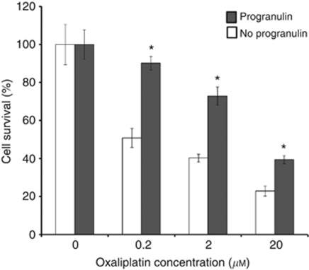
Background: Recent studies suggest that cancer stem cells (CSCs) mediate chemoresistance, but interestingly, only a small percentage of cells in a resistant tumour are CSCs; this suggests that non-CSCs survive by other means. We hypothesised that chemoresistant colorectal cancer (CRC) cells generate soluble factors that enhance survival of chemonaive tumour cells.

Methods: Chemoresistant CRC cells were generated by serial passage in oxaliplatin (Ox cells). Conditioned media (CM) was collected from parental and oxaliplatin-resistant (OxR) cells. CRC cells were treated with CM and growth and survival were assessed. Tumour growth rates were determined in nude mice after cells were treated with CM. Mass spectrometry (MS) identified proteins in CM. Reverse phase protein microarray assays determined signalling effects of CM in parental cells.

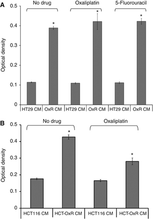
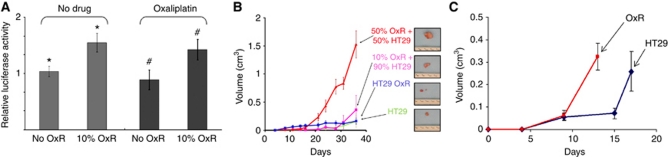
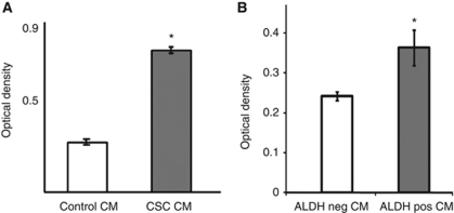
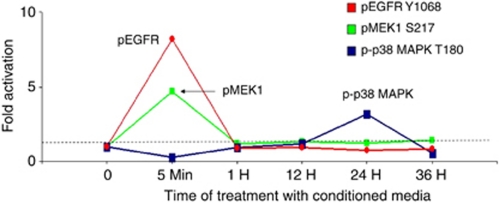
Results: Oxaliplatin-resistant CM increased survival of chemo-naive cells. CSC CM also increased growth of parental cells. Parental and OxR mixed tumours grew larger than tumours composed of parental or OxR cells alone. Mass spectrometry detected unique survival-promoting factors in OxR CM compared with parental CM. Cells treated with OxR CM demonstrated early phosphorylation of EGFR and MEK1, with later upregulation of total Akt .We identified progranulin as a potential mediator of chemoresistance.

Conclusion: Chemoresistant tumour cells and CSCs may promote resistance through soluble factors that mediate survival in otherwise chemosensitive tumour cells.


Figures

Figure 1
Figure 1
Effect of CM from OxR cells on CRC cell proliferation and survival. (A) Proliferative effect of CM from OxR HT29 cells. MTT assay on HT29 cells treated with CM from parental HT29 cells (HT29 CM) or from OxR HT29 cells (OxR CM). Cells were either untreated or treated with oxaliplatin as described in the Materials and Methods section. The y-axis displays OD readings from a plate reader as values relative to blank wells containing DMSO only. P-value <0.001.(P-values were calculated by a two-tailed Student's t-test for significance). (B) Proliferative effect of CM from OxR HCT116 cells. MTT assay on HCT116 cells treated with CM from parental HCT116 cells (HCT116 CM) or from OxR HCT116 cells (HCT-OxR CM). Cells were either untreated or treated with oxaliplatin as described in the Materials and Methods section. *P-value <0.001.
Figure 2
Figure 2
Effect of OxR cells in mixed cultures and mixed tumours. (A) In vitro mixed-cell experiment. Luciferase activity of ‘bystander’ parental HT29 cells when cultured alone (no OxR cells) or when mixed with 10% OxR cells, either untreated or treated with oxaliplatin. Data are normalised to the control set (no OxR cells). P-values were calculated by a two-tailed Student's t-test for significance. The symbols * and # each indicate P=0.03 (B) In vivo mixed-tumour experiment. Growth curves of in vivo tumours composed of four ratios of cells as indicated, with representative photographs of tumours from each cohort. The y-axis represents volumes as calculated from caliper measurements. (C) Growth curves of secondary tumours in mice with parental HT29 (blue line) and OxR (red line) feeder tumours implanted in the contralateral flank. Days indicate time elapsed after subcutaneous injection of secondary tumours. Feeder tumour sizes of the HT29- and OxR-derived tumours were 0.15 and 0.04 cm3 on day 0, respectively, and reached 2.43 and 0.17 cm3, respectively, at the termination of the experiment.
Figure 3
Figure 3
Effect of CM from cultures enriched for CSCs on crc cell proliferation and survival. (A) Effect of CM from side-population-based CSCs on growth of HT29 parental cells. Conditioned media were obtained from parental HT29 (control CM) or cells enriched for the CSC phenotype by the side-population assay. These media were used to treat HT29 cells for 48 h, and then MTT assays were performed on triplicate cultures. (B) Effect of conditioned media from Aldefluor-based enrichment for CSCs on growth of HT29 parental cells. Conditioned media were obtained from HT29 sorted by the Aldefluor assay for Aldefluor-negative (ALDH neg CM) and Aldefluor-positive (ALDH pos CM) cells. These media were used to treat HT29 cells for 48 h, and then MTT assays were performed on triplicate cultures. *P<0.05.
Figure 4
Figure 4
Effect of CM from OxR cells on signalling pathways in target cells: pathway activation mapping analysis. HT29 cells were treated with CM from parental HT29 cells and OxR HT29 cells for times indicated and then analysed by reverse-phase protein microarray. Values are expressed as fold change of protein activation/phosphorylation or expression, as indicated, of cells treated with CM from OxR cells vs parental cells. Phosphorylation of EGFR, MEK1, and p38 MAPK are depicted by red, green, and blue graphs, respectively.
Figure 5
Figure 5
Effect of progranulin on sensitivity to chemotherapy of HT29 colorectal cancer cells treated with oxaliplatin. HT29 cells were treated with or without 1 μg ml−1 of recombinant progranulin and oxaliplatin at the indicated concentrations. Cell survival/proliferation was then assayed by MTT assay. Data bars represent the ratio of optical densities of oxaliplatin-treated cells to untreated cells (0) at 96 h for cells treated with progranulin (shaded bars) or cells not treated with progranulin (empty bars). *P<0.05.

References

    1. Arumugam T, Ramachandran V, Logsdon CD (2006) Effect of cromolyn on S100P interactions with RAGE and pancreatic cancer growth and invasion in mouse models. J Natl Cancer Inst 98: 1806–1818 - PMC - PubMed
    1. Barker N, Ridgway RA, van Es JH, van de Wetering M, Begthel H, van den Born M, Danenberg E, Clarke AR, Sansom OJ, Clevers H (2009) Crypt stem cells as the cells-of-origin of intestinal cancer. Nature 457: 608–611 - PubMed
    1. Beatty GL, Giantonio BJ (2008) Bevacizumab and oxaliplatin-based chemotherapy in metastatic colorectal cancer. Expert Rev Anticancer Ther 8: 683–688 - PubMed
    1. Bhushan S, McLeod H, Walko CM (2009) Role of pharmacogenetics as predictive biomarkers of response and/or toxicity in the treatment of colorectal cancer. Clin Colorectal Cancer 8: 15–21 - PubMed
    1. Blazer III DG, Kishi Y, Maru DM, Kopetz S, Chun YS, Overman MJ, Fogelman D, Eng C, Chang DZ, Wang H, Zorzi D, Ribero D, Ellis LM, Glover KY, Wolff RA, Curley SA, Abdalla EK, Vauthey JN (2008) Pathologic response to preoperative chemotherapy: a new outcome end point after resection of hepatic colorectal metastases. J Clin Oncol 26: 5344–5351 - PubMed

Publication types

MeSH terms