Review
. 2011 Nov 1;8(1):43-51.
doi: 10.1038/nrneph.2011.168.

The pathogenesis and diagnosis of acute kidney injury in multiple myeloma

Affiliations
Review

The pathogenesis and diagnosis of acute kidney injury in multiple myeloma

Colin A Hutchison et al. Nat Rev Nephrol. .

Abstract

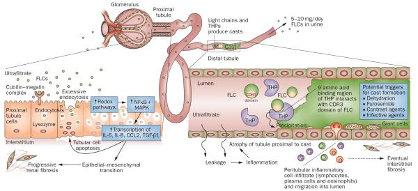
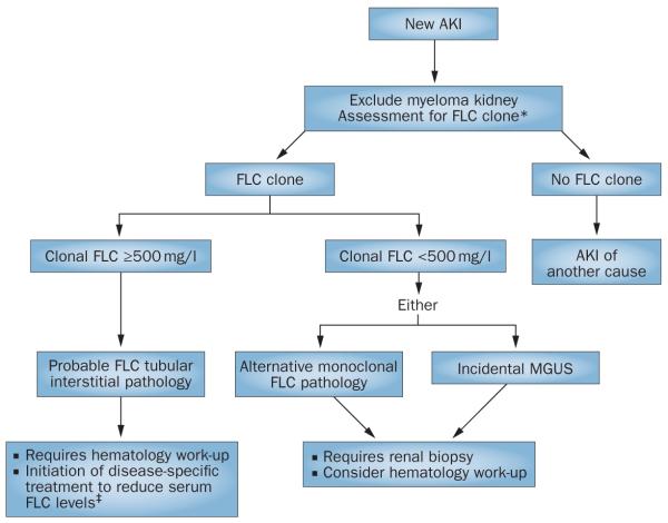
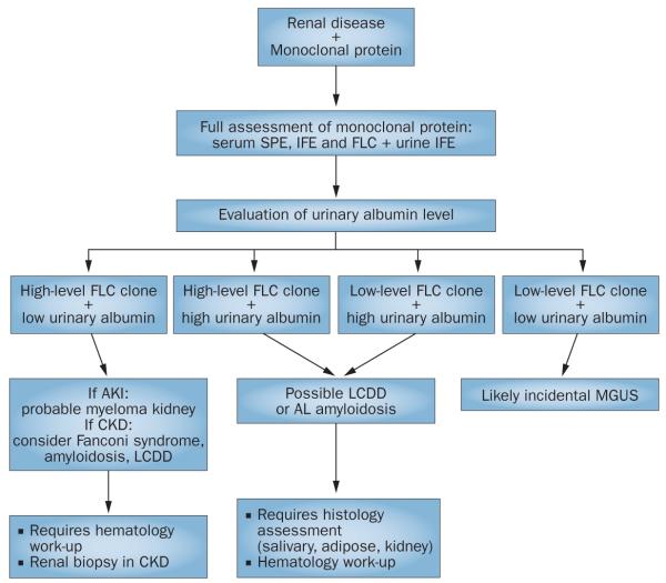
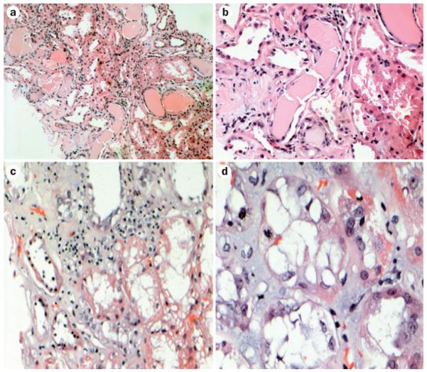
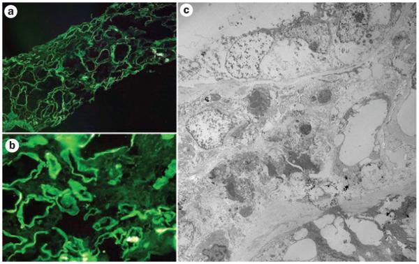
Renal failure remains a principal cause of morbidity for patients with multiple myeloma. Once reversible factors such as hypercalcemia have been corrected, the most common cause of severe renal failure in these patients is a tubulointerstitial pathology that results from the very high circulating concentrations of monoclonal immunoglobulin free light chains. These endogenous proteins can result in isolated proximal tubule cell cytotoxicity, tubulointerstitial nephritis and cast nephropathy (myeloma kidney). Less frequently, high levels of free light chains can lead to immunoglobulin light chain amyloidosis and light chain deposition disease, although these conditions are usually associated with insidious progression of renal failure rather than acute kidney injury. Unless there is rapid intervention, progressive and irreversible damage occurs, particularly interstitial fibrosis and tubular atrophy. Despite advances in our understanding of the pathogenesis of these processes there has been a gap in translating these achievements into improved patient outcomes. The International Kidney and Monoclonal Gammopathy Research Group was formed to address this need. In this Review, we discuss the mechanisms of disease and diagnostic approaches to patients with acute kidney injury complicating multiple myeloma.


Figures

Figure 1
Figure 1
Mechanisms of FLC-induced acute kidney injury. The very high concentrations of FLCs present in the ultrafiltrate of patients with multiple myeloma can result in direct injury to PTCs. The excessive endocytosis of FLCs by the cubilin–megalin complex expressed on PTCs can trigger apoptotic, proinflammatory and fibrotic pathways. Activation of redox pathways occurs, with increased expression of NFκB and MAPK, which in turn leads to the transcription of both inflammatory and profibrotic cytokines, such as IL-6, IL-8, CCL2 and TGF-β1. In the distal tubules, FLCs can bind to a specific binding domain on THPs and co-precipitate to form casts. These casts result in tubular atrophy proximal to the cast and lead to progressive interstitial inflammation and fibrosis. Abbreviations: CCL2, C-C motif chemokine 2; CDR, complementarity determining region; FLC, free light chain; IL, interleukin; MAPK, mitogen-activated protein kinase; NFκB, nuclear factor κB; PTC, proximal tubule cell; TGF-β1, transforming growth factor β1; THP, Tamm-Horsfall protein.
Figure 2
Figure 2
Screening algorithm for monoclonal disease in AKI. Patients with AKI can be initially screened for a monoclonal protein by serum FLC assays, which enable the rapid identification of a monoclonal FLC as the possible cause of a tubular interstitial process. *To exclude the presence of an intact monoclonal immunoglobin, serum FLC assays should be combined with serum protein electrophoresis (many units would also include immunofixation electrophoresis as standard of care). For the assessment of immunoglobulin light chain amyloidosis and light chain deposition disease, urinary assessment is required. Where serum FLC assays are not used, urgent urinary assessment for monoclonal FLCs is necessary. Emergency treatment designed to achieve a rapid reduction in serum FLC levels is based on high-dose dexamethasone. The full benefit of adding FLC removal strategies to this therapy remains to be determined. Abbreviations: AKI, acute kidney injury; FLC, free light chain; MGUS, monoclonal gammopathy of undetermined significance.
Figure 3
Figure 3
Diagnostic approach to a patient with renal disease and a monoclonal protein. Combining the evaluation of urinary albumin concentration with the level of the FLC clone can guide the management of a patient with renal injury and a monoclonal protein. Tubulointerstitial pathologies are more likely when urinary albumin levels are low and FLC levels are high. By contrast, patients with AL amyloidosis and LCDD frequently have high urinary albumin levels. These pathologies are often associated with a lower level of FLC clone, but can occur with any FLC level. Where diagnostic uncertainty remains, assessment of histology is essential. Abbreviations: AKI, acute kidney injury; AL, amyloid light chain; CKD, chronic kidney disease; FLC, free light chain; IFE, immunofixation electrophoresis; LCDD, light chain deposition disease; MGUS, monoclonal gammopathy of undetermined significance; SPE, serum protein electrophoresis.
Figure 4
Figure 4
Renal injuries induced by free light chains in patients with acute kidney injury. All samples were stained with hematoxylin and eosin. The classic appearance of cast nephropathy can be seen in a | and b | (magnification ×150 and ×350, respectively). Casts with fracture planes in distal nephrons and surrounding reactive tubular cells can be seen (center). Note adjacent proximal tubular damage and mild inflammation surrounding the casts. c | Marked tubular damage is observed, with inflammatory infiltrate that contains a mixture of mononuclear inflammatory cells and scattered eosinophils (magnification ×250). d | Tubular interstitial injury without casts (magnification ×750). A mitotic figure can be seen in a tubular cell, indicative of tubular regeneration.
Figure 5
Figure 5
Immunofluorescence and electron microscopy evaluation of tubulointerstitial damage associated with circulating monoclonal free light chains. a | and b | show examples of immunofluorescence for κ light chains. Linear staining along the tubular basement membranes represents κ light chains (negative for λ light chains). c | Electron microscopy findings of prominent tubular damage and interstitial inflammatory infiltrate. Focal punctate electron-dense material around tubular basement membranes represents deposits of free light chains.

References

    1. Peto R. Factors of prognostic significance in myelomatosis. J. Clin. Pathol. 1972;25:555. - PMC - PubMed
    1. Haynes RJ, Read S, Collins GP, Darby SC, Winearls CG. Presentation and survival of patients with severe acute kidney injury and multiple myeloma: a 20-year experience from a single centre. Nephrol. Dial. Transplant. 2010;25:419–426. - PubMed
    1. Bladé J, et al. Renal failure in multiple myeloma: presenting features and predictors of outcome in 94 patients from a single institution. Arch. Intern. Med. 1998;158:1889–1893. - PubMed
    1. Leung N, et al. Improvement of cast nephropathy with plasma exchange depends on the diagnosis and on reduction of serum free light chains. Kidney Int. 2008;73:1282–1288. - PubMed
    1. Hutchison CA, et al. Treatment of acute renal failure secondary to multiple myeloma with chemotherapy and extended high cut-off hemodialysis. Clin. J. Am. Soc. Nephrol. 2009;4:745–754. - PMC - PubMed

Publication types

MeSH terms