Comparative Study
. 2012 Jan 18;31(2):457-70.
doi: 10.1038/emboj.2011.384. Epub 2011 Nov 1.

Palmitoylated TMX and calnexin target to the mitochondria-associated membrane

Affiliations
Comparative Study

Palmitoylated TMX and calnexin target to the mitochondria-associated membrane

Emily M Lynes et al. EMBO J. .

Abstract

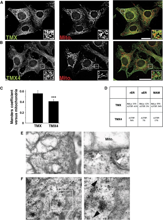
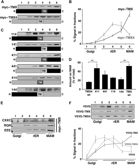
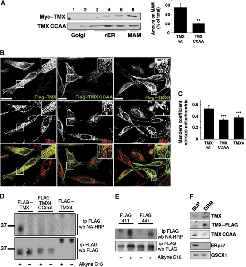
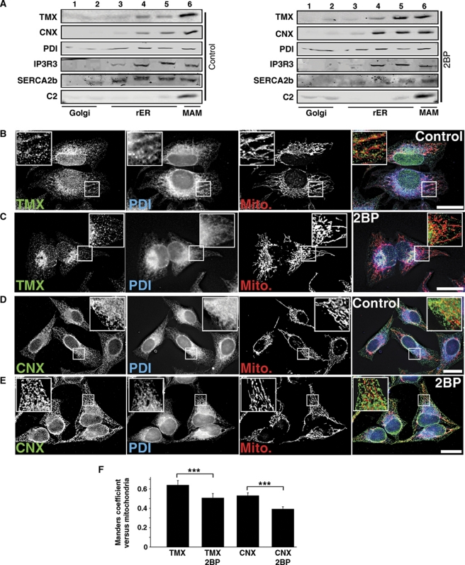
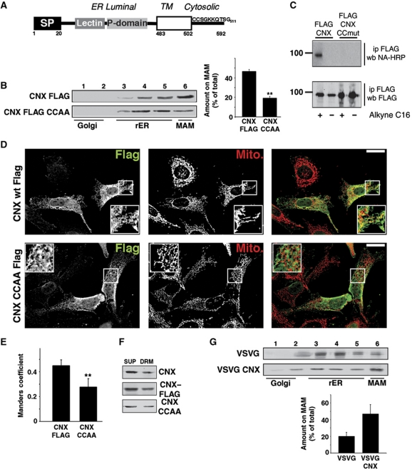
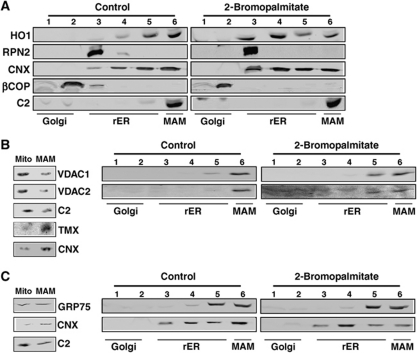
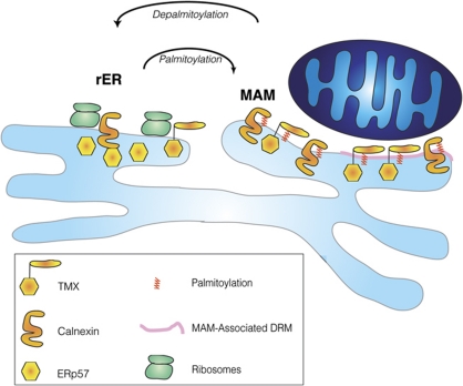
The mitochondria-associated membrane (MAM) is a domain of the endoplasmic reticulum (ER) that mediates the exchange of ions, lipids and metabolites between the ER and mitochondria. ER chaperones and oxidoreductases are critical components of the MAM. However, the localization motifs and mechanisms for most MAM proteins have remained elusive. Using two highly related ER oxidoreductases as a model system, we now show that palmitoylation enriches ER-localized proteins on the MAM. We demonstrate that palmitoylation of cysteine residue(s) adjacent to the membrane-spanning domain promotes MAM enrichment of the transmembrane thioredoxin family protein TMX. In addition to TMX, our results also show that calnexin shuttles between the rough ER and the MAM depending on its palmitoylation status. Mutation of the TMX and calnexin palmitoylation sites and chemical interference with palmitoylation disrupt their MAM enrichment. Since ER-localized heme oxygenase-1, but not cytosolic GRP75 require palmitoylation to reside on the MAM, our findings identify palmitoylation as key for MAM enrichment of ER membrane proteins.


Conflict of interest statement

The authors declare that they have no conflict of interest.

Figures

Figure 1
Figure 1
The thioredoxin-related transmembrane protein TMX co-fractionates with the MAM in A375P and HeLa cells. (A) Diagram of the topology of TMX proteins. Indicated are signal peptides (SP), thioredoxin domains (TRX), and the transmembrane domains (open boxes). Positions of these domains and of the CXXC active sites are indicated with numbers. (B) Analysis of expression levels of TMX family proteins. Lysates from HeLa, A375P, Jurkat, Caco2, and M2 melanoma cells were analysed by western blot for the respective oxidoreductases using α-tubulin as a protein loading control. Molecular weight markers are on the left. (C) Analysis of the ER domain distribution of TMX family proteins in A375P and HeLa cells. Cell homogenates were fractionated on an Optiprep gradient. eIF2α indicates the position of the rER (fractions 3–5), βCOP indicates the cis-Golgi (fraction 2), the peak of the calnexin signal indicates the MAM (fraction 6). (D) Quantification of the fractionation of TMX and TMX4. Three individual experiments in HeLa and A375P cells, respectively, were quantified. The graph shows the distribution of TMX and TMX4 within all fractions, in addition to the respective amounts within fraction 6. (Statistics: ***P=0.0026 between HeLa TMX and TMX4, P=0.001 between A375P TMX and TMX4.) (E) TMX and calnexin distribution between mitochondria and the MAM. HeLa homogenates were fractionated into cytosol (Cyt.), microsomes (Micro.), crude mitochondria (MC), purified mitochondria (MP), and MAM according to Materials and methods. Equal cell equivalents have been loaded.
Figure 2
Figure 2
Intracellular localization of endogenous TMX and TMX4. (A) HeLa cells were processed for immunofluorescence microscopy for TMX and mitochondria (visualized with Mitotracker). Images were deconvolved. Inserts show a magnified area, indicated with white frames on the bigger pictures. Yellow colour in the overlap indicates apposition of the TMX and mitochondria staining. Scale bar=25 μm. A representative image is shown. (B) A375P cells were processed and imaged as in (A) using a mouse TMX4 antibody and mitotracker. (C) Quantification of the Manders coefficient for the localization of TMX and TMX4, as shown by representative images in (A, B). (Statistics: ***P<0.001.) (D) Quantification of TMX and TMX4 immunogold labelling from two independent experiments with the localization of TMX and TMX4 assigned to rER, sER, and MAM (229 particles for TMX in HeLa, 174 particles for TMX4 in A375P). (E) HeLa cells were treated for TMX immunogold labelling as described in Materials and methods. A representative image is shown to the left with a zoomed-in area on the right. In all, 10 nm anti-rabbit immunogold particles are highlighted with white arrowheads. Scale bar=200 nm. (F) A375P cells were treated for TMX4 immunogold labelling as described in Materials and methods, a representative image is shown with a zoomed-in area. In all, 10 nm anti-mouse immunogold particles are highlighted with black arrowheads. Scale bar=200 nm.
Figure 3
Figure 3
The TMX transmembrane and cytosolic domains are necessary and sufficient for MAM targeting. Except for (F), detection of tagged proteins with a polyclonal anti-myc antiserum. (A) Optiprep gradient fractionation of luminally myc-tagged TMX and TMX4. HeLa cells transfected with myc–TMX and myc–TMX4 were fractionated on an Optiprep gradient, reproducing the fractionation pattern as seen for endogenous counterparts. (B) Quantification of the Optiprep fractionation of myc–TMX and myc–TMX4. Positions of marker proteins are indicated (n=3). (C) Optiprep gradient analysis of TMX/TMX4 chimera. HeLa cells transfected with TMX/TMX4 chimera were analysed as in (A). Chimera has been assigned a code, where 1 stands for TMX and 4 for TMX4 as follows: luminal/transmembrane domain/cytosolic domain. (D) Quantification of the Optiprep fractionation for selected TMX/TMX4 chimera. Coding as in (C). (Statistics: **P=0.027 between TMX4 wt and 411; P=0.012 between TMX wt and 144.) (E) Optiprep gradient fractionation for selected point mutants. Amino acids indicated stand for alanines that substitute them. CXXC=Δ54–63; RQR=Arg, Glu, Arg267–269 Ala; EEE=Asp248–250 → Ala. (F) Optiprep gradient fractionation of VSVG-TMX/TMX4 chimera. HeLa cells transfected with wild-type VSVG and chimera of luminal VSVG fused to transmembrane and cytosolic domains of TMX and TMX4, respectively, were fractionated on an Optiprep gradient. Detection by western blotting with a rabbit anti-VSVG antiserum (n=3).
Figure 4
Figure 4
TMX contains two cytosolic, transmembrane-proximal cysteine residues that mediate MAM enrichment. (A) Mutagenesis of cysteines 205 and 207 abrogates MAM enrichment of myc-tagged TMX. HeLa cells transfected with myc–TMX and myc–TMX (Cys205, 207 → Ala) were fractionated on a discontinuous 10–30% Optiprep gradient, and enrichment of these mutants on the MAM was determined by western blot. A quantification of three independent experiments is shown on the right. (Statistics: **P=0.012 between wt and CCAA TMX.) (B) Immunofluorescence localization of FLAG-tagged TMX, TMX CCAA, and TMX4. HeLa cells were transfected for 8 h with the indicated constructs and processed for immunofluorescence microscopy for the FLAG tag and mitochondria (visualized with Mitotracker). Images were deconvolved. Inserts show a magnified area, indicated with white frames on the bigger pictures. Yellow colour in the overlap indicates apposition of the TMX and mitochondria staining. Scale bar=25 μm. Representative images are shown. (C) Quantification of the Manders coefficient for the localization of the constructs, as shown by representative images in (E). (Statistics: ***P<0.001.) (D) Detection of palmitoylation on TMX and TMX4 constructs. Top gel shows the alkynyl-palmitate detection with HRP-conjugated neutravidin. Bottom gel shows the input, as evidenced by the anti-FLAG western blot signal. Size markers are shown on the left. Efficient palmitoylation is detected on FLAG-tagged TMX only. (E) Detection of palmitoylation on TMX chimera (labelled as in C). Top gel shows the alkynyl-palmitate detection with HRP-conjugated neutravidin. Bottom gel shows the input, as evidenced by the anti-FLAG western blot signal. (F) Detection of TMX on DRMs. DRM-associated proteins (DRM) were separated from detergent-soluble supernatants (SUP). Proteins analysed (top to bottom): endogenous TMX, transfected FLAG-tagged TMX, transfected FLAG-tagged TMX CCAA (palmitoylation-deficient mutant). ERp57 and QSOX1 were used as controls.
Figure 5
Figure 5
2-Bromopalmitate reduces the apposition of the TMX and calnexin signals with mitochondria. (A) Block of palmitoylation with 2-bromopalmitate abrogates MAM enrichment of endogenous TMX and calnexin. HeLa cells were fractionated on an Optiprep gradient, and enrichment of TMX and calnexin on the MAM was determined by western blot. The ER proteins PDI, IP3R3, SERCA2b and the mitochondrial complex 2 serve as controls for MAM integrity. (B) HeLa cells were processed for immunofluorescence microscopy for TMX, PDI, and mitochondria (visualized with Mitotracker). Images were deconvolved. Inserts show a magnified area, indicated with white frames on the bigger pictures. Scale bar=25 μm. A representative image is shown. (C) 2-Bromopalmitate-treated HeLa cells were processed for immunofluorescence microscopy as in (B). (D) HeLa cells were processed for immunofluorescence microscopy for calnexin, PDI, and mitochondria (visualized with Mitotracker). Images were deconvolved. Inserts show a magnified area, indicated with white frames on the bigger pictures. Scale bar=25 μm. A representative image is shown. (E) 2-Bromopalmitate-treated HeLa cells were processed for immunofluorescence microscopy as in (D). (F) Quantification of the Manders coefficient for the localization of TMX and calnexin, as shown by representative images in (BE). (Statistics: ***P<0.001 as indicated.)
Figure 6
Figure 6
Palmitoylation is necessary and sufficient for calnexin MAM enrichment. (A) Diagram of the topology of human calnexin. Indicated are signal peptide (SP), the lectin and P domain, and the transmembrane domain (open box). Positions of selected domains are indicated with numbers. (B) Mutagenesis of transmembrane-proximal cysteines 503/504 to alanines abrogates MAM enrichment of FLAG-tagged calnexin. Control HeLa cells and HeLa cells transfected with FLAG–calnexin and FLAG–calnexin (Cys503, 504 → Ala) were fractionated on an Optiprep gradient, and enrichment of these proteins on the MAM was determined by western blot. A quantification of three independent experiments is shown on the right. (Statistics: **P=0.002 between CNX wt and CNX CCAA.) (C) Detection of palmitoylation on FLAG-tagged calnexin. Top gel shows the alkynyl-palmitate detection with HRP-conjugated neutravidin. Bottom gel shows the input, as evidenced by the anti-FLAG western blotting signal. Size markers are shown on the left. (D) Immunofluorescence localization of FLAG-tagged calnexin and calnexin CCAA. HeLa cells were transfected for 8 h with the indicated constructs and processed for immunofluorescence microscopy for the FLAG tag and mitochondria (visualized with Mitotracker). Images were deconvolved. Inserts show a magnified area, indicated with white frames on the bigger pictures. Yellow colour in the overlap indicates apposition of the calnexin and mitochondria staining. Scale bar=25 μm. Representative images are shown. (E) Quantification of the Manders coefficient for the localization of the constructs, as shown by representative images in (E). (Statistics: **P=0.0044.) (F) Detection of calnexin on DRMs. DRM-associated proteins (DRM) were separated from detergent-soluble supernatants (SUP). Proteins analysed (top to bottom): endogenous calnexin, transfected FLAG-tagged calnexin, transfected FLAG-tagged calnexin CCAA (palmitoylation-deficient mutant). (G) Optiprep gradient fractionation of VSVG-calnexin chimera. HeLa cells transfected with wild-type VSVG and chimera of luminal VSVG fused to the transmembrane and cytosolic domain of calnexin were fractionated on an Optiprep gradient. Detection by western blot with a rabbit anti-VSVG antiserum (n=3).
Figure 7
Figure 7
2-Bromopalmitate reduces MAM enrichment of heme oxygenase-1, but not of VDAC1/2 and GRP75. (A) Block of palmitoylation with 2-bromopalmitate abrogates MAM enrichment of heme oxygenase-1. B16 cells were fractionated on a discontinuous 10–30% Optiprep gradient, and enrichment of heme oxygenase-1 on the MAM was determined by western blot. The ER proteins ribophorin-2, and calnexin, as well as βCOP and mitochondrial complex 2 serve as controls for MAM integrity. (B) VDAC1 and VDAC2 show a 2-bromopalmitate-sensitive moiety. Left gels: Percoll separation of mitochondria and MAM. VDAC1/2 show a mitochondrial distribution, when compared with MAM proteins TMX and calnexin. Right gels: Block of palmitoylation with 2-bromopalmitate shifts a moiety of VDAC1/2 into rER fractions. HeLa cells were fractionated on an Optiprep gradient, and enrichment of VDAC1/2 on the MAM was determined by western blot. (C) GRP75 is not mis-localized with 2-bromopalmitate. Left gels: Percoll separation of mitochondria and MAM. GRP75 localizes in part to MAM. Right gels: Block of palmitoylation with 2-bromopalmitate does not affect GRP75. HeLa cells were fractionated on an Optiprep gradient, and enrichment of GRP75 on the MAM was determined by western blot.
Figure 8
Figure 8
Model for palmitoylation-dependent MAM enrichment of ER chaperones and oxidoreductases. Palmitoylated calnexin and TMX (indicated with the red modifications) target to the MAM, which is composed of DRM (pink domain) and non-DRM.

References

    1. Abrami L, Kunz B, Iacovache I, van der Goot FG (2008) Palmitoylation and ubiquitination regulate exit of the Wnt signaling protein LRP6 from the endoplasmic reticulum. Proc Natl Acad Sci USA 105: 5384–5389 - PMC - PubMed
    1. Anelli T, Alessio M, Bachi A, Bergamelli L, Bertoli G, Camerini S, Mezghrani A, Ruffato E, Simmen T, Sitia R (2003) Thiol-mediated protein retention in the endoplasmic reticulum: the role of ERp44. EMBO J 22: 5015–5022 - PMC - PubMed
    1. Anelli T, Bergamelli L, Margittai E, Rimessi A, Fagioli C, Malgaroli A, Pinton P, Ripamonti M, Rizzuto R, Sitia R (2011) Ero1alpha regulates Ca2+ fluxes at the endoplasmic reticulum-mitochondria interface (MAM). Antioxid Redox Signal (in press) - PubMed
    1. Appenzeller-Herzog C, Ellgaard L (2008) The human PDI family: versatility packed into a single fold. Biochim Biophys Acta 1783: 535–548 - PubMed
    1. Area-Gomez E, de Groof AJ, Boldogh I, Bird TD, Gibson GE, Koehler CM, Yu WH, Duff KE, Yaffe MP, Pon LA, Schon EA (2009) Presenilins are enriched in endoplasmic reticulum membranes associated with mitochondria. Am J Pathol 175: 1810–1816 - PMC - PubMed

Publication types

MeSH terms